
Why does 10% fetal bovine serum drastically reduce the inhibitory potency of Rapamycin?
Quick Answer
Recommended ApproachAssay rapamycin potency under low-serum conditions (0.1–1% FBS) or serum-starved baselines, adjusting working concentrations to compensate for serum protein sequestration and mitogen-driven mTORC1 reactivation.
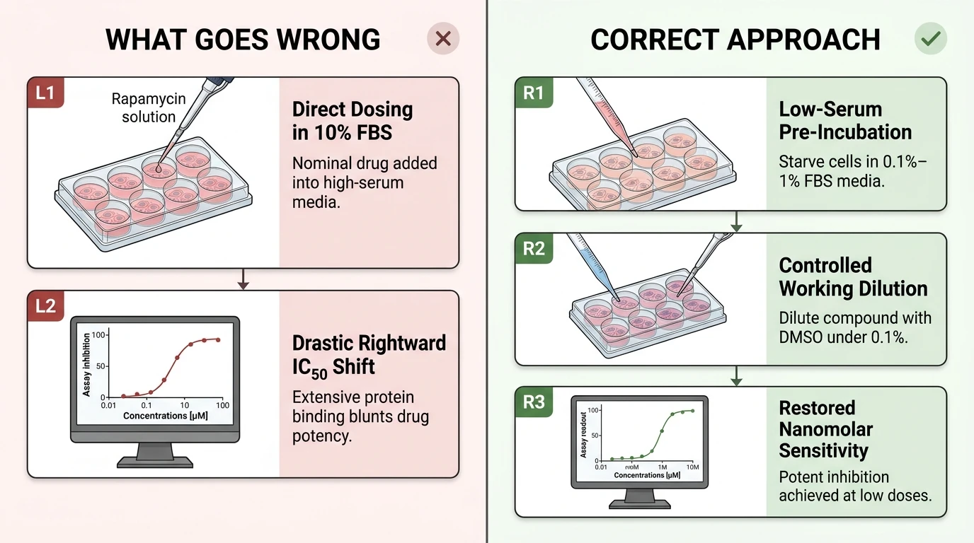
Common CausesTesting rapamycin in standard 10% FBS culture media dramatically elevates the apparent IC50 because serum albumin and alpha-1-acid glycoprotein extensively sequester lipophilic rapamycin, while abundant growth factors hyperactivate upstream PI3K/Akt/mTOR signaling to override allosteric mTORC1 suppression.
How Should It Be Done Correctly?
Copy code to add this image to your page Copy Code
In Vitro Assay Parameters for Rapamycin Potency
Recommended culture and dosing parameters for evaluating rapamycin inhibition across serum conditions.
Stepwise Assay Protocol for High-Sensitivity mTORC1 Inhibition
- Stock Reconstitution and Aliquoting: Dissolve rapamycin powder (MW 914.18) in anhydrous DMSO to prepare a 10 mM stock solution. Aliquot into single-use, low-binding amber tubes to avoid repeated freeze-thaw cycles, storing long-term at -80°C (1-year shelf life) or short-term at -20°C (1-month shelf life).
- Cell Seeding and Density Optimization: Seed adherent target cells into multi-well plates and allow 16–24 hours for attachment until reaching 60%–70% confluence. Avoid over-confluency, as contact inhibition alters basal mTOR and Akt phosphorylation levels.
- Serum Adaptation and Starvation Step: Decant full-growth media (10% FBS) and wash cell monolayers gently with pre-warmed PBS. Replace with reduced-serum media (0.1%–0.5% FBS) or serum-free basal medium for 4 to 16 hours. This step stabilizes basal phosphorylation of mTOR, p70S6K, and 4E-BP1 to establish a reproducible baseline [2, 4].
- Serial Dilution Preparation: Prepare serial dilutions of rapamycin in serum-free medium immediately before treatment. Maintain identical DMSO concentrations across all drug dilutions and the vehicle control, ensuring the final organic solvent concentration remains at or below 0.1% (v/v) [4].
- Compound Incubation and Pathway Evaluation: Apply the compound dilutions to cells and incubate for 1 to 24 hours depending on the target readout (acute signaling vs. anti-proliferative phenotype) [3, 4, 5]. Lyse cells directly on ice with cold lysis buffer supplemented with protease and phosphatase inhibitors to preserve phosphorylation states [4, 5].
| Parameter | Standard Serum Culture | Low-Serum Potency Assay | Serum-Deprived Starvation | Recommended Stock Solution |
|---|---|---|---|---|
| Serum Concentration | 10% FBS | 0.1%–1% FBS | 0% FBS (serum-free DMEM) | N/A (neat anhydrous DMSO) |
| Rapamycin Working Range | 10 nM–10 µM [1, 2] | 0.5 nM–100 nM [3] | 0.1 nM–20 nM [2, 4] | 10 mM in DMSO |
| Free (Unbound) Fraction | < 1%–5% | ~20%–50% | ~100% | N/A |
| Pre-incubation Starvation | None | 4–16 h before compound addition | 1–24 h prior to readout [4] | N/A |
| Target Biomarkers | p-mTOR, p-p70S6K, p-4E-BP1 [1, 2] | p-S6K1, p-Akt Ser473 [5] | p-mTOR, p-p70S6K, LC3B-II [4] | LC3-II autophagic flux [4] |
| Vehicle (DMSO) Limit | ≤ 0.1% (v/v) [4] | ≤ 0.1% (v/v) | ≤ 0.1% (v/v) | Store stock at -80°C (1 yr) or -20°C (1 mo) |
What Went Wrong?
Uncorrected Compound Addition into High-Serum Media
Assaying rapamycin potency directly in regular growth medium containing 10% fetal bovine serum creates an apparent rightward shift in the half-maximal inhibitory concentration (IC50), blunting both target dephosphorylation and downstream antiproliferative responses [1, 2]. Investigators frequently mistake this marked drop in biological potency for compound degradation or cellular resistance, compensating by escalating doses up into micromolar ranges where off-target or incomplete responses emerge [2, 3].
Depletion of Bioavailable Free Drug by Serum Proteins
Rapamycin is a highly lipophilic macrolide lactone that displays extensive, high-affinity non-specific binding to serum proteins, particularly bovine serum albumin (BSA) and lipoproteins. In 10% FBS, the vast majority of rapamycin molecules are partitioned into the protein-bound fraction, drastically depleting the concentration of free, uncomplexed drug available to cross cellular membranes and bind its cytosolic receptor. Consequently, the effective intracellular dose is only a small fraction of the nominal concentration added to the well.
Hyperactive Upstream Mitogenic Drive and Allosteric Resistance
Fetal bovine serum contains abundant growth factors (e.g., IGF-1, EGF, PDGF) and amino acids that activate receptor tyrosine kinases, driving the PI3K/Akt signaling axis to sustain maximal mTORC1 kinase activity [2, 4]. Mechanistically, rapamycin acts not as a catalytic ATP-competitive inhibitor, but as an allosteric inhibitor: it must first form a gain-of-function binary complex with FK506-binding protein 12 (FKBP12) before binding the FRB domain of mTOR, inducing dissociation of Raptor from mTORC1 [6, 7]. In the presence of 10% FBS: - Persistent mitogen stimulation drives redundant hyperphosphorylation of p70S6K and 4E-BP1, rendering specific mTORC1 substrates (especially 4E-BP1 Thr37/46) relatively refractory to rapamycin-mediated shutdown [1, 2]. - Starvation or serum deprivation starkly sensitizes mTORC1 to rapamycin by eliminating competing upstream inputs, allowing complete inhibition of downstream translation and induction of autophagy at sub-nanomolar to low-nanomolar concentrations [2, 4]. - Prolonged allosteric mTORC1 blockage in nutrient-rich conditions triggers feedback activation of upstream Akt through the loss of p70S6K-mediated IRS-1 degradation, sustaining cell survival and masking rapamycin efficacy in long-term viability assays [2, 5].
How Can I Avoid This in Future Experiments?
To decouple serum interference from true pharmacological potency, calibrate assay concentrations against the specific serum content of the media. When testing in full-serum media (10% FBS) is necessary to sustain cell viability or mimic in vivo conditions, anticipate a 10- to 100-fold increase in the apparent IC50 relative to serum-free or low-serum conditions [1, 2, 3]. Alternatively, utilize a brief serum-starvation period (2–4 hours) followed by acute drug pre-treatment before pulsing with serum or defined growth factors; this reveals the unencumbered inhibitory capacity of rapamycin against downstream effectors such as p70S6K and 4E-BP1.
References
- [1]Pu BP, et al. World journal of gastrointestinal oncology. 2025 Sep;17(9):109378.[IF: 2.7]
- [2]Ruffolo B, et al. Cancers. 2025 Aug;17(15).[IF: 4.8]
- [3]Asani B, et al. BMC ophthalmology. 2022 Mar;22(1):138.[IF: 2.0]
- [4]Ga YJ, et al. Pharmaceuticals (Basel, Switzerland). 2025 Dec;18(12).[IF: 5.7]
- [5]Romanyuk N, et al. Frontiers in cellular neuroscience. 2024 Jan;18:1298182.[IF: 4.0]
- [6]Dai X, et al. Scientific reports. 2024 Jan;14(1):2082.[IF: 3.9]
- [7]Abdelrahman SA, et al. Iranian journal of basic medical sciences. 2022 Jun;25(6):732-744.[IF: 2.2]