-
 Australia
-
 Austria
-
 Belgium
-
 Brazil
-
 Canada
-
 China
China
-
 Czech Republic
-
 Denmark
-
 Finland
-
 France
-
 Germany
-
 Greece
-
 Hong Kong
-
 Hungary
-
 Iceland
-
 India
India
-
 Ireland
-
 Israel
-
 Italy
Italy
-
 Japan
Japan
-
 Korea
Korea
-
 Luxembourg
-
 Malaysia
-
 Netherlands
-
 New Zealand
-
 Norway
-
 Poland
-
 Qatar
Qatar
-
 Romania
-
 Saudi Arabia
-
 Singapore
-
 Spain
Spain
-
 Sweden
-
 Switzerland
-
 Taiwan
-
 Turkey
-
 United Kingdom
-
 United States
research use only
Mdivi-1 Drp1 inhibitor
Cat.No.S7162
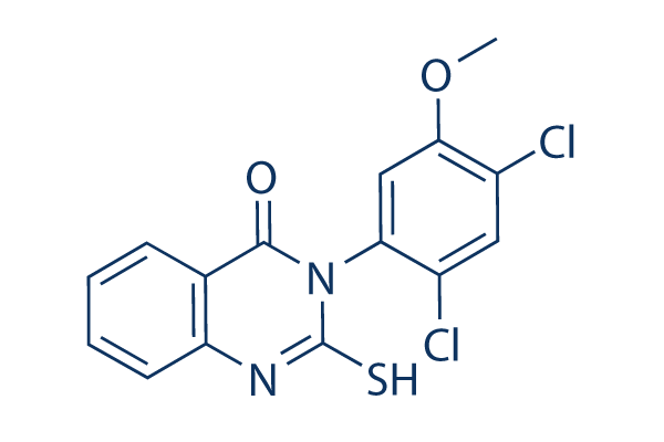
Chemical Structure
Molecular Weight: 353.22
Quality Control
| Related Targets | Akt Wnt/beta-catenin PKC HSP ROCK Microtubule Associated Integrin Bcr-Abl Actin FAK |
|---|---|
| Other Dynamin Inhibitors | Dynasore Dyngo-4a (Hydroxy-Dynasore) P110 MiTMAB TPH104m Dynole 34-2 |
Cell Culture, Treatment & Working Concentration
| Cell Lines | Assay Type | Concentration | Incubation Time | Formulation | Activity Description | PMID |
|---|---|---|---|---|---|---|
| L1210 | Function assay | 2.5, 5 and 10 μM | 72 h | Mdivi-1 (5 µM) significantly attenuated 0.4 mg/l of cell death in L1210 cells, with the exception of 2.5 and 10 µM of Mdivi-1 | 28677814 | |
| INS-1E | Function assay | 50 μM | 12 h | treatment with Mdivi-1 significantly inhibited mitochondrial fragmentation and clearly increased the viabilityofpancreatic beta INS-1E cells under hypoxia conditions | 29768513 | |
| R28 | Function assay | 5 μM | 2 h | mitochondria in R28 cells pretreated with 5 μM Drp1 inhibitor Mdivi-1 retained the average length and appeared as elongated tubular, thread-like networks after irradiation | 30538621 | |
| HepG2/ADM cells | Cell viability assay | 10 μM | 1 h | mdivi-1 pretreatment increased B5G1-induced apoptosis and cell death in HepG2/ADM cells | 30850585 | |
| SH-SY5Y | Function assay | 10 μM | 30 min | Inhibiting Drp1 protects against CPF-induced intrinsic apoptosis by blocking Bax translocation | 26598294 | |
| Click to View More Cell Line Experimental Data | ||||||
Solubility
|
In vitro |
DMSO
: 125 mg/mL
(353.88 mM)
Water : Insoluble Ethanol : Insoluble |
Molarity Calculator
|
In vivo |
|||||
In vivo Formulation Calculator (Clear solution)
Step 1: Enter information below (Recommended: An additional animal making an allowance for loss during the experiment)
Step 2: Enter the in vivo formulation (This is only the calculator, not formulation. Please contact us first if there is no in vivo formulation at the solubility Section.)
Calculation results:
Working concentration: mg/ml;
Method for preparing DMSO master liquid: mg drug pre-dissolved in μL DMSO ( Master liquid concentration mg/mL, Please contact us first if the concentration exceeds the DMSO solubility of the batch of drug. )
Method for preparing in vivo formulation: Take μL DMSO master liquid, next addμL PEG300, mix and clarify, next addμL Tween 80, mix and clarify, next add μL ddH2O, mix and clarify.
Method for preparing in vivo formulation: Take μL DMSO master liquid, next add μL Corn oil, mix and clarify.
Note: 1. Please make sure the liquid is clear before adding the next solvent.
2. Be sure to add the solvent(s) in order. You must ensure that the solution obtained, in the previous addition, is a clear solution before proceeding to add the next solvent. Physical methods such
as vortex, ultrasound or hot water bath can be used to aid dissolving.
Chemical Information, Storage & Stability
| Molecular Weight | 353.22 | Formula | C15H10Cl2N2O2S |
Storage (From the date of receipt) | |
|---|---|---|---|---|---|
| CAS No. | 338967-87-6 | Download SDF | Storage of Stock Solutions |
|
|
| Synonyms | Mitochondrial division inhibitor 1 | Smiles | COC1=C(C=C(C(=C1)N2C(=O)C3=CC=CC=C3NC2=S)Cl)Cl | ||
Mechanism of Action
| Features |
The first selective inhibitor of mitochondrial division dynamins.
|
|---|---|
| Targets/IC50/Ki |
Dnm1 GTPase
(Cell-free assay) 1 μM-10 μM
|
| In vitro |
Mdivi-1 is a cell-permeable quinazolinone compound that inhibits yeast (Dnm1) and mammalian (Drp1) division DRPs (dynamin-related GTPases) and effectively induces mitochondrial fusion into net-like structures in a reversible manner. Cell-free studies indicate that this compound blocks Dnm1 ATPase activity (IC50<10 μM) and self-assembly by an allosteric modulation-based mechanism. It is shown to effectively suppress STS- as well as C8-Bid-induced MOMP (Mitochondrial Outer Membrane Permeabilization) in HeLa cultures and in cell-free murine liver mitochondria preparations, respectively, as assessed by cytochrome C release. In cells, this chemical retards apoptosis by inhibiting mitochondrial outer membrane permeabilization. In principle, this compound represents a class of therapeutics for stroke, myocardial infarction, and neurodegenerative diseases. |
| In vivo |
Drp1 and GFAP protein expression is significantly increased in the early neurodegenerative events of ischemic mouse retina. Mdivi-1 treatment blocks apoptotic cell death in ischemic retina, and significantly increases RGC survival at 2 weeks after ischemia. In the normal mouse retina, Drp1 is expressed in the ganglion cell layer (GCL) as well as the inner plexiform layer, the inner nuclear layer (INL), and the outer plexiform layer (OPL). In the GCL, Drp1 immunoreactivity is strong in RGCs. While Drp1 protein expression is increased in the GCL of vehicle-treated ischemic retina at 12 hours. This compound treatment does not change this increase of Drp1 protein expression but significantly decreased GFAP protein expression. |
References |
|
Applications
| Methods | Biomarkers | Images | PMID |
|---|---|---|---|
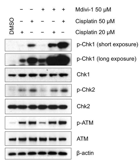
| Western blot | p-Chk1 / p-Chk2 / Chk1 / Chk2 / p-ATM / ATM COX-2 / p-Drp1 / Drp1 / Mfn2 / ABCG2 / Oct4 |
|
24952704 |
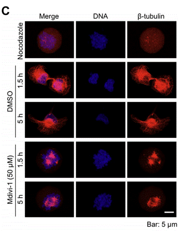
| Immunofluorescence | β-tubulin |
|
25458053 |
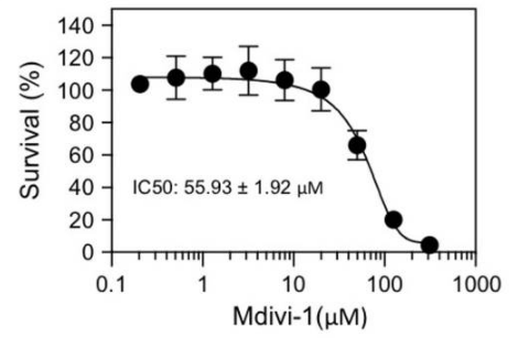
| Growth inhibition assay | Cell viability Apoptosis |
|
24952704 |
Tech Support
Tel: +1-832-582-8158 Ext:3
If you have any other enquiries, please leave a message.
Products are for research use only. Not for human use. We do not sell to patients.
©Copyright 2013 Selleck Chemicals. All Rights Reserved.