-
 Australia
-
 Austria
-
 Belgium
-
 Brazil
-
 Canada
-
 China
China
-
 Czech Republic
-
 Denmark
-
 Finland
-
 France
-
 Germany
-
 Greece
-
 Hong Kong
-
 Hungary
-
 Iceland
-
 India
India
-
 Ireland
-
 Israel
-
 Italy
Italy
-
 Japan
Japan
-
 Korea
Korea
-
 Luxembourg
-
 Malaysia
-
 Netherlands
-
 New Zealand
-
 Norway
-
 Poland
-
 Qatar
Qatar
-
 Romania
-
 Saudi Arabia
-
 Singapore
-
 Spain
Spain
-
 Sweden
-
 Switzerland
-
 Taiwan
-
 Turkey
-
 United Kingdom
-
 United States
research use only
Pyroxamide (NSC 696085) HDAC inhibitor
Cat.No.S2190
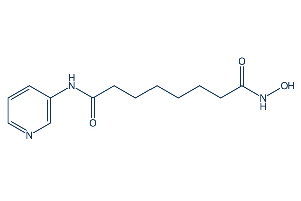
Chemical Structure
Molecular Weight: 265.31
Quality Control
Solubility
|
In vitro |
DMSO
: 17 mg/mL
(64.07 mM)
Ethanol : 2 mg/mL Water : Insoluble |
Molarity Calculator
|
In vivo |
|||||
In vivo Formulation Calculator (Clear solution)
Step 1: Enter information below (Recommended: An additional animal making an allowance for loss during the experiment)
Step 2: Enter the in vivo formulation (This is only the calculator, not formulation. Please contact us first if there is no in vivo formulation at the solubility Section.)
Calculation results:
Working concentration: mg/ml;
Method for preparing DMSO master liquid: mg drug pre-dissolved in μL DMSO ( Master liquid concentration mg/mL, Please contact us first if the concentration exceeds the DMSO solubility of the batch of drug. )
Method for preparing in vivo formulation: Take μL DMSO master liquid, next addμL PEG300, mix and clarify, next addμL Tween 80, mix and clarify, next add μL ddH2O, mix and clarify.
Method for preparing in vivo formulation: Take μL DMSO master liquid, next add μL Corn oil, mix and clarify.
Note: 1. Please make sure the liquid is clear before adding the next solvent.
2. Be sure to add the solvent(s) in order. You must ensure that the solution obtained, in the previous addition, is a clear solution before proceeding to add the next solvent. Physical methods such
as vortex, ultrasound or hot water bath can be used to aid dissolving.
Chemical Information, Storage & Stability
| Molecular Weight | 265.31 | Formula | C13H19N3O3 |
Storage (From the date of receipt) | |
|---|---|---|---|---|---|
| CAS No. | 382180-17-8 | Download SDF | Storage of Stock Solutions |
|
|
Mechanism of Action
| Targets/IC50/Ki |
HDAC1
100 nM(ID50)
|
|---|---|
| In vitro |
Pyroxamide (NSC 696085) causes the accumulation of acetylated core histones in MEL cells cultured with the agent. At micromolar concentrations, it induces terminal differentiation and inhibits proliferation of murine erythroleukemia(MEL) cells. This compound (1.25-20 mM) causes dose-dependent inhibition of growth by cell cycle arrest of prostate carcinoma (LNCaP), neuroblastoma (KCN-69n), and bladder carcinoma (T24) cells in culture, with similar efficacy in all of the cell lines. A concentration of 1.25-20.0 μM Pyroxamide causes a dose-dependent decrease in viable cell number and an increase in percentage of dead cells over time in two Rhabdomyosarcoma cell lines, RD (embryonal ) and RH30B (alveolar). Accumulation of acetylated histones and induction of p21/WAF1 expression are obersevd in cells exposed to it. It shows a dose- and time-dependent proliferation inhibition, induction of apoptosis and histone H4 hyperacetylation in three B-cell precursor (BCP)-acute lymphoblastic leukemia (ALL) cell lines (Reh, Nalm6, Z33). The calculated IC50 after 96 hours of its incubation are 2-6 μM. |
| Kinase Assay |
HDAC Inhibition Assays
|
|
A MEL cell line expressing the epitope Flag-tagged HDAC1 is generated. HDAC1-Flag is affinity purified by immune-precipitation using M2 anti-Flag antibody-coated agarose, followed by elution from the agarose using the Flag peptide. [3H]acetate-labeled cellular histones are prepared from MEL cells and are used as a substrate for the HDAC activity assay. Released [3H] acetic acid is quantified by scintillation counting. For inhibition studies, the enzyme preparations are preincubated with Pyroxamide (NSC 696085) (10 to 100,000 nM) for 30 minutes at 4 °
|
|
| In vivo |
Administration of 100 or 200 mg/kg of Pyroxamide (NSC 696085) daily for 21 days causes significant, dose-dependent suppression of the growth of the tumor xenograft. A dose of 300 mg/kg of this compound is lethal to all of the mice in the treatment group within 1 week. After in vivo administration, accumulation of acetylated histones and a dose-dependent increase in the expression of p21/WAF1 protein level are observed. |
References |
|
Tech Support
Tel: +1-832-582-8158 Ext:3
If you have any other enquiries, please leave a message.
Signaling Pathway Map
Products are for research use only. Not for human use. We do not sell to patients.
©Copyright 2013 Selleck Chemicals. All Rights Reserved.