-
 Australia
-
 Austria
-
 Belgium
-
 Brazil
-
 Canada
-
 China
China
-
 Czech Republic
-
 Denmark
-
 Finland
-
 France
-
 Germany
-
 Greece
-
 Hong Kong
-
 Hungary
-
 Iceland
-
 India
India
-
 Ireland
-
 Israel
-
 Italy
Italy
-
 Japan
Japan
-
 Korea
Korea
-
 Luxembourg
-
 Malaysia
-
 Netherlands
-
 New Zealand
-
 Norway
-
 Poland
-
 Qatar
Qatar
-
 Romania
-
 Saudi Arabia
-
 Singapore
-
 Spain
Spain
-
 Sweden
-
 Switzerland
-
 Taiwan
-
 Turkey
-
 United Kingdom
-
 United States
research use only
PCI-34051 HDAC8 Inhibitor
Cat.No.S2012
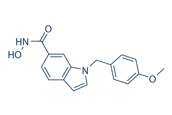
Chemical Structure
Molecular Weight: 296.32
Quality Control
Cell Culture, Treatment & Working Concentration
| Cell Lines | Assay Type | Concentration | Incubation Time | Formulation | Activity Description | PMID |
|---|---|---|---|---|---|---|
| LAN1 | Growth inhibition assay | 72 hrs | Growth inhibition of human LAN1 cells incubated for 72 hrs by MTS assay, GI50=3.9μM | 23116147 | ||
| Jurkat | Growth inhibition assay | 72 hrs | Growth inhibition of human Jurkat cells incubated for 72 hrs by MTS assay, GI50=11μM | 23116147 | ||
| NB-1 | Growth inhibition assay | 72 hrs | Growth inhibition of human NB-1 cells incubated for 72 hrs by MTS assay, GI50=14μM | 23116147 | ||
| MT2 | Growth inhibition assay | 72 hrs | Growth inhibition of human MT2 cells incubated for 72 hrs by MTS assay, GI50=15μM | 23116147 | ||
| MT4 | Growth inhibition assay | 72 hrs | Growth inhibition of human MT4 cells incubated for 72 hrs by MTS assay, GI50=25μM | 23116147 | ||
| Huh7 | Antiviral assay | 3 days | Antiviral activity against HCV genotype 1b infected in human Huh7 cells after 3 days by luciferase reporter gene assay, EC50=1.8μM | 25490700 | ||
| HuH7 | Cytotoxicity assay | 3 days | Cytotoxicity against human HuH7 cells assessed as inhibition of cell viability after 3 days by CellTiter 96 assay, CC50=11μM | 25490700 | ||
| Sf9 | Function assay | 5 mins | Inhibition of recombinant full-length human C-terminal FLAG-His-tagged HDAC1 expressed in baculovirus infected Sf9 insect cells using Boc-L-Lys(Ac)-AMC as substrate preincubated for 5 mins followed by substrate addition measured after 35 mins by fluoresce, IC50=3.8μM | 28835796 | ||
| Sf9 | Function assay | 5 mins | Inhibition of recombinant human full-length C-terminal His-tagged HDAC3 (395 to 489 residues)/human NCOR2 expressed in baculovirus infected Sf9 insect cells using Boc-L-Lys(Ac)-AMC as substrate pretreated for 5 mins followed by substrate addition measured, IC50=7.1μM | 28835796 | ||
| Sf9 | Function assay | 5 mins | Inhibition of recombinant human full-length C-terminal His-tagged HDAC2 expressed in baculovirus infected Sf9 insect cells using Boc-L-Lys(Ac)-AMC as substrate preincubated for 5 mins followed by substrate addition measured after 35 mins by fluorescence assay, IC50=31μM | 28835796 | ||
| SH-SY5Y | Cytotoxicity assay | Cytotoxicity against human SH-SY5Y cells expressing TP53 by CellTiter96 AQueous one solution cell proliferation assay | 28835796 | |||
| IMR5 | Cytotoxicity assay | Cytotoxicity against human IMR5 cells expressing TP53 by CellTiter96 AQueous one solution cell proliferation assay | 28835796 | |||
| SK-N-AS | Cytotoxicity assay | Cytotoxicity against human SK-N-AS cells expressing TP53 mutation by CellTiter96 AQueous one solution cell proliferation assay | 28835796 | |||
| Kelly | Cytotoxicity assay | Cytotoxicity against human Kelly cells expressing TP53 mutation by CellTiter96 AQueous one solution cell proliferation assay | 28835796 | |||
| BE(2)-C | Cytotoxicity assay | 72 hrs | Cytotoxicity against human BE(2)-C cells assessed as reduction in cell viability by measuring metabolic activity after 72 hrs by WST-8 assay, IC50=19.9μM | 29190092 | ||
| Sf9 | Function assay | 90 mins | Inhibition of recombinant human full length C-terminal FLAG-tagged HDAC1 expressed in fall armyworm Sf9 cells using fluorogenic ZMAL as substrate after 90 mins by fluorimetric analysis, IC50=28.3μM | 29190092 | ||
| Sf9 | Function assay | 90 mins | Inhibition of recombinant human full length HDAC6 expressed in fall armyworm Sf9 cells using fluorogenic ZMAL as substrate after 90 mins by fluorimetric analysis, IC50=48.2μM | 29190092 | ||
| BE(2)-C | Function assay | 6 uM | 72 hrs | Inhibition of HDAC8 in human BE(2)-C cells assessed as upregulation of p21/CDKN1 gene expression at 6 uM after 72 hrs by RT-PCR analysis relative to control | 29190092 | |
| BE(2)-C | Function assay | 6 uM | 72 hrs | Inhibition of HDAC8 in human BE(2)-C cells assessed as upregulation of TrkA/NTRK1 gene expression at 6 uM after 72 hrs by RT-PCR analysis relative to control | 29190092 | |
| BE(2)-C | Function assay | 6 uM | 72 hrs | Inhibition of HDAC8 in human BE(2)-C cells assessed as upregulation of TH gene expression at 6 uM after 72 hrs by RT-PCR analysis relative to control | 29190092 | |
| BE(2)-C | Function assay | 6 uM | 6 days | Induction of outgrowth of neurofilament positive neutrite-like structures in human BE(2)-C cells at 6 uM after 6 days by DAPI-staining based microscopic analysis | 29190092 | |
| A673 | qHTS assay | qHTS of pediatric cancer cell lines to identify multiple opportunities for drug repurposing: Primary screen for A673 cells | 29435139 | |||
| SK-N-MC | qHTS assay | qHTS of pediatric cancer cell lines to identify multiple opportunities for drug repurposing: Primary screen for SK-N-MC cells | 29435139 | |||
| NB-EBc1 | qHTS assay | qHTS of pediatric cancer cell lines to identify multiple opportunities for drug repurposing: Primary screen for NB-EBc1 cells | 29435139 | |||
| SK-N-SH | qHTS assay | qHTS of pediatric cancer cell lines to identify multiple opportunities for drug repurposing: Primary screen for SK-N-SH cells | 29435139 | |||
| NB1643 | qHTS assay | qHTS of pediatric cancer cell lines to identify multiple opportunities for drug repurposing: Primary screen for NB1643 cells | 29435139 | |||
| LAN-5 | qHTS assay | qHTS of pediatric cancer cell lines to identify multiple opportunities for drug repurposing: Primary screen for LAN-5 cells | 29435139 | |||
| Jurkat | Antiproliferative assay | Antiproliferative activity against human Jurkat cells by alamar blue assay, GI50=2.4μM | 29505935 | |||
| HUT78 | Antiproliferative assay | Antiproliferative activity against human HUT78 cells by alamar blue assay, GI50=2.4μM | 29505935 | |||
| HSB2 | Antiproliferative assay | Antiproliferative activity against human HSB2 cells by alamar blue assay, GI50=2.4μM | 29505935 | |||
| MOLT4 | Antiproliferative assay | Antiproliferative activity against human MOLT4 cells by alamar blue assay, GI50=2.4μM | 29505935 | |||
| Jurkat | Antiproliferative assay | 48 hrs | Antiproliferative activity against human Jurkat cells after 48 hrs by MTT assay, IC50=4.5μM | 29533873 | ||
| MOLT4 | Antiproliferative assay | 48 hrs | Antiproliferative activity against human MOLT4 cells after 48 hrs by MTT assay, IC50=9.4μM | 29533873 | ||
| HEL | Antiproliferative assay | 48 hrs | Antiproliferative activity against human HEL cells after 48 hrs by MTT assay, IC50=10.8μM | 29533873 | ||
| SK-N-BE(2) | Antiproliferative assay | 48 hrs | Antiproliferative activity against human SK-N-BE(2) cells after 48 hrs by MTT assay, IC50=16.9μM | 29533873 | ||
| PC3 | Antiproliferative assay | 48 hrs | Antiproliferative activity against human PC3 cells after 48 hrs by MTT assay, IC50=19.2μM | 29533873 | ||
| K562 | Antiproliferative assay | 72 hrs | Antiproliferative activity against human K562 cells after 72 hrs by MTS assay, GI50=2.01μM | 30004697 | ||
| K562R | Antiproliferative assay | 72 hrs | Antiproliferative activity against human K562R cells after 72 hrs by MTS assay, GI50=2.2μM | 30004697 | ||
| HCT116 | Antiproliferative assay | 72 hrs | Antiproliferative activity against human HCT116 cells after 72 hrs by MTS assay, GI50=2.64μM | 30004697 | ||
| PC3 | Antiproliferative assay | 72 hrs | Antiproliferative activity against human PC3 cells after 72 hrs by MTS assay, GI50=2.66μM | 30004697 | ||
| MCF7 | Antiproliferative assay | 72 hrs | Antiproliferative activity against human MCF7 cells after 72 hrs by MTS assay, GI50=2.97μM | 30004697 | ||
| BL21 (DE3) | Function assay | Binding affinity to human His-thioredoxin-tagged HDAC8 expressed in Escherichia coli BL21 (DE3) cells by ITC method, Kd=0.0751μM | 30347148 | |||
| BL21 (DE3) | Function assay | Inhibition of human His-thioredoxin-tagged HDAC8 expressed in Escherichia coli BL21 (DE3) cells using Fluor de Lys (R)-HDAC8 as substrate by fluorometric method, IC50=0.0777μM | 30347148 | |||
| BL21 (DE3) | Function assay | Binding affinity to Schistosoma mansoni His-tagged HDAC8 expressed in Escherichia coli BL21 (DE3) cells by ITC method, Kd=0.367μM | 30347148 | |||
| BL21 (DE3) | Function assay | Inhibition of Schistosoma mansoni His-tagged HDAC8 expressed in Escherichia coli BL21 (DE3) cells using Fluor de Lys (R)-HDAC8 as substrate by fluorometric method, IC50=0.4358μM | 30347148 | |||
| BL21 (DE3) | Function assay | Inhibition of human His-thioredoxin-tagged HDAC8 mL6/L179I mutant expressed in Escherichia coli BL21 (DE3) cells using Fluor de Lys (R)-HDAC8 as substrate by fluorometric method, IC50=1μM | 30347148 | |||
| BL21 (DE3) | Function assay | Inhibition of human His-thioredoxin-tagged HDAC8 mL6 mutant expressed in Escherichia coli BL21 (DE3) cells using Fluor de Lys (R)-HDAC8 as substrate by fluorometric method, IC50=1.63μM | 30347148 | |||
| BL21 (DE3) | Function assay | Inhibition of human His-thioredoxin-tagged HDAC8 mL1/mL6 mutant expressed in Escherichia coli BL21 (DE3) cells using Fluor de Lys (R)-HDAC8 as substrate by fluorometric method, IC50=2.7μM | 30347148 | |||
| BL21 (DE3) | Function assay | Inhibition of human His-thioredoxin-tagged HDAC8 mL1/mL6/L179I mutant expressed in Escherichia coli BL21 (DE3) cells using Fluor de Lys (R)-HDAC8 as substrate by fluorometric method, IC50=5μM | 30347148 | |||
| Sf9 | Function assay | Inhibition of C-terminal FLAG/His-tagged full length human HDAC1 expressed in baculovirus infected Sf9 insect cells using Z(Ac)Lys-AMC as substrate by fluorometric method, IC50=28.3μM | 30347148 | |||
| Sf9 | Function assay | Inhibition of N-terminal GST-tagged full length human HDAC6 expressed in baculovirus infected Sf9 insect cells using Z(Ac)Lys-AMC as substrate by fluorometric method, IC50=48.2μM | 30347148 | |||
| Sf9 | Function assay | 40 mins | Inhibition of recombinant human full length C-terminal His-tagged HDAC8 expressed in baculovirus infected insect cells measured after 40 mins by HDAC-Glo1/2 luminescent assay, IC50=0.03162μM | 30964290 | ||
| SK-N-BE(2)C | Anticlonogenic assay | 96 hrs | Anticlonogenic activity in human SK-N-BE(2)C cells assessed as reduction in cell proliferation incubated for 96 hrs by crystal violet staining based assay, GI50=15μM | 31630054 | ||
| Click to View More Cell Line Experimental Data | ||||||
Solubility
|
In vitro |
DMSO
: 59 mg/mL
(199.1 mM)
Water : Insoluble Ethanol : Insoluble |
Molarity Calculator
|
In vivo |
|||||
In vivo Formulation Calculator (Clear solution)
Step 1: Enter information below (Recommended: An additional animal making an allowance for loss during the experiment)
Step 2: Enter the in vivo formulation (This is only the calculator, not formulation. Please contact us first if there is no in vivo formulation at the solubility Section.)
Calculation results:
Working concentration: mg/ml;
Method for preparing DMSO master liquid: mg drug pre-dissolved in μL DMSO ( Master liquid concentration mg/mL, Please contact us first if the concentration exceeds the DMSO solubility of the batch of drug. )
Method for preparing in vivo formulation: Take μL DMSO master liquid, next addμL PEG300, mix and clarify, next addμL Tween 80, mix and clarify, next add μL ddH2O, mix and clarify.
Method for preparing in vivo formulation: Take μL DMSO master liquid, next add μL Corn oil, mix and clarify.
Note: 1. Please make sure the liquid is clear before adding the next solvent.
2. Be sure to add the solvent(s) in order. You must ensure that the solution obtained, in the previous addition, is a clear solution before proceeding to add the next solvent. Physical methods such
as vortex, ultrasound or hot water bath can be used to aid dissolving.
Chemical Information, Storage & Stability
| Molecular Weight | 296.32 | Formula | C17H16N2O3 |
Storage (From the date of receipt) | |
|---|---|---|---|---|---|
| CAS No. | 950762-95-5 | Download SDF | Storage of Stock Solutions |
|
|
| Synonyms | N/A | Smiles | COC1=CC=C(C=C1)CN2C=CC3=C2C=C(C=C3)C(=O)NO | ||
Mechanism of Action
| Targets/IC50/Ki |
HDAC8
(Cell-free assay) 10 nM
|
|---|---|
| In vitro |
PCI-34051 possesses promising potency for HDAC8 with a Ki of 10 nM. This compound has high selectivity (approximately fivefold) for HDAC8 relative to the other class I HDACs including HDAC1. It reveals greater than 200-fold selectivity over HDAC1 and HDAC6, and greater than 1000-fold selectivity over HDAC2, HDAC3 and HDAC10. This chemical inhibits ovarian tumor line OVCAR-3 with a GI50 of 6 μM and 15% cell death. Neither significant tubulin nor histone acetylation is observed in the sensitive cell lines treated with this compound at concentrations less than 25 μM at 24 hours nor at earlier timepoints. It induces a selective cytotoxic effect in cell lines derived only from T-cell malignancies. This compound induces caspase-dependent apoptosis. When caspase-3 activity is measured at various times after treatment with 5 μM of this chemical, increasing levels of activity are observed from 12 to 24 to 48 hours, another hallmark of apoptosis, consistent with the higher levels of caspase activity at this timepoint. It does not stimulate Bid cleavage, a characteristic effect of the extrinsic apoptotic pathway. While P116 and J.RT3-T.5 are sensitive to this compound, the PLCγ1-deficient J.gamma1 line reveals a marked decrease in the extent of its induced apoptosis. In addition, steady-state calcium levels strongly influence the apoptosis induced by this chemical. It induces cytochrome c release from mitochondria. |
| Kinase Assay |
Histone deacetylase activity
|
|
For PCI-34051 characterization, measurements are perfomed in a reaction volume of 100 μL using 96-well assay plates in a fluorescence plate reader. For each isozyme. The HDAC protein in reaction buffer (50 mM HEPES, 100 mM KCl, 0.001% Tween-20, 5% dimethyl sulfoxide, pH7.4, supplemented with bovine serum albumin at concentrations of 0-0.05%) is mixed with this compound at various concentrations and allowed to incubate for 15 min. Trysin is added to a final concentration of 50 nM, and acetyl-gly-Ala-(N-acetyl-Lys)-amino-4-methylcoumarin is added to a final concentration of 25-100 μM to initiate the reaction. After a 30 min lag time, the fluorescence is measured over a 30 min time frame using an excitation wavelength of 335 nm and a detection wavelength of 460 nm. The increase in fluorescence wih time is used as the measure of the reaction rate.
|
|
| In vivo |
PCI-34051 is a potent and specific HDAC8 inhibitor. |
References |
|
Applications
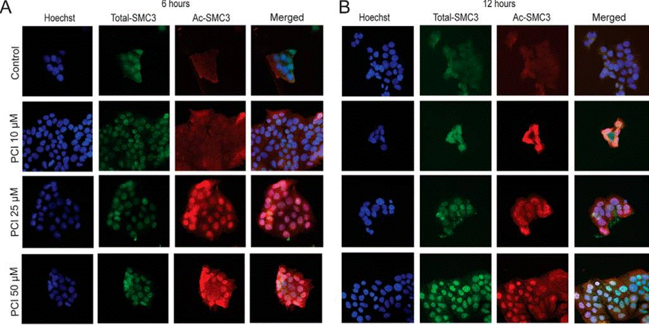
| Methods | Biomarkers | Images | PMID |
|---|---|---|---|
| Immunofluorescence | SMC3 / Ac-SMC3 |
|
27072133 |
Tech Support
Tel: +1-832-582-8158 Ext:3
If you have any other enquiries, please leave a message.
Signaling Pathway Map
Products are for research use only. Not for human use. We do not sell to patients.
©Copyright 2013 Selleck Chemicals. All Rights Reserved.