-
 Australia
-
 Austria
-
 Belgium
-
 Brazil
-
 Canada
-
 China
China
-
 Czech Republic
-
 Denmark
-
 Finland
-
 France
-
 Germany
-
 Greece
-
 Hong Kong
-
 Hungary
-
 Iceland
-
 India
India
-
 Ireland
-
 Israel
-
 Italy
Italy
-
 Japan
Japan
-
 Korea
Korea
-
 Luxembourg
-
 Malaysia
-
 Netherlands
-
 New Zealand
-
 Norway
-
 Poland
-
 Qatar
Qatar
-
 Romania
-
 Saudi Arabia
-
 Singapore
-
 Spain
Spain
-
 Sweden
-
 Switzerland
-
 Taiwan
-
 Turkey
-
 United Kingdom
-
 United States
research use only
TCS7010 (Aurora A Inhibitor I) Aurora Kinase inhibitor
Cat.No.S1451
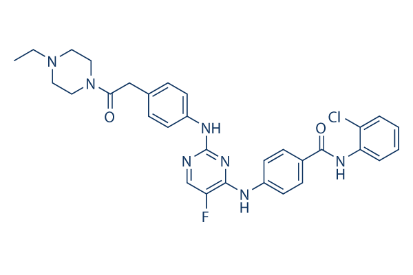
Chemical Structure
Molecular Weight: 588.07
Quality Control
| Related Targets | CDK HSP PD-1/PD-L1 ROCK Wee1 DNA/RNA Synthesis Microtubule Associated Ras KRas Casein Kinase |
|---|---|
| Other Aurora Kinase Inhibitors | Hesperadin Barasertib-HQPA (AZD2811) Alisertib (MLN8237) Tozasertib (VX-680, MK-0457) ZM 447439 MLN8054 Danusertib (PHA-739358) MK-5108 AMG-900 CCT137690 |
Cell Culture, Treatment & Working Concentration
| Cell Lines | Assay Type | Concentration | Incubation Time | Formulation | Activity Description | PMID |
|---|---|---|---|---|---|---|
| HCT116 cells | Proliferation assay | 4 days | Antiproliferative activity against human HCT116 cells after 4 days by celltiter assay, IC50=0.19 μM | |||
| HT-29 cells | Proliferation assay | 4 days | Antiproliferative activity against human HT-29 cells after 4 days by celltiter assay, IC50=2.9 μM | |||
| Click to View More Cell Line Experimental Data | ||||||
Solubility
|
In vitro |
DMSO
: 25 mg/mL
(42.51 mM)
Water : Insoluble Ethanol : Insoluble |
Molarity Calculator
|
In vivo |
|||||
In vivo Formulation Calculator (Clear solution)
Step 1: Enter information below (Recommended: An additional animal making an allowance for loss during the experiment)
Step 2: Enter the in vivo formulation (This is only the calculator, not formulation. Please contact us first if there is no in vivo formulation at the solubility Section.)
Calculation results:
Working concentration: mg/ml;
Method for preparing DMSO master liquid: mg drug pre-dissolved in μL DMSO ( Master liquid concentration mg/mL, Please contact us first if the concentration exceeds the DMSO solubility of the batch of drug. )
Method for preparing in vivo formulation: Take μL DMSO master liquid, next addμL PEG300, mix and clarify, next addμL Tween 80, mix and clarify, next add μL ddH2O, mix and clarify.
Method for preparing in vivo formulation: Take μL DMSO master liquid, next add μL Corn oil, mix and clarify.
Note: 1. Please make sure the liquid is clear before adding the next solvent.
2. Be sure to add the solvent(s) in order. You must ensure that the solution obtained, in the previous addition, is a clear solution before proceeding to add the next solvent. Physical methods such
as vortex, ultrasound or hot water bath can be used to aid dissolving.
Chemical Information, Storage & Stability
| Molecular Weight | 588.07 | Formula | C31H31ClFN7O2 |
Storage (From the date of receipt) | |
|---|---|---|---|---|---|
| CAS No. | 1158838-45-9 | Download SDF | Storage of Stock Solutions |
|
|
| Synonyms | TC-S 7010 | Smiles | CCN1CCN(CC1)C(=O)CC2=CC=C(C=C2)NC3=NC=C(C(=N3)NC4=CC=C(C=C4)C(=O)NC5=CC=CC=C5Cl)F | ||
Mechanism of Action
| Features |
Aurora A Inhibitor I is a novel, potent, and selective inhibitor to Aurora A.
|
|---|---|
| Targets/IC50/Ki |
Aurora A
(Cell-free assay) 3.4 nM
|
| In vitro |
TCS7010 (Aurora A Inhibitor I) is a 2,4-dianilinopyrimidine that selectively and potently inhibits Aurora A. It effectively inhibits the proliferation of HCT116 and HT29 cells, with IC50 of 190 nM and 2.9 μM, respectively. The Aurora A selectivity of this compound against Aurora B depends on a single amino acid (Thr217) of Aurora A. In KCL-22 cells, it (1-5 μM) increases G2/M cell fraction, induces histone H3 serine 10 phosphorylation, and suppresses mitotic Aurora A autophosphorylation on Thr288. It (0.5-5 μM) also suppresses cell proliferation in KCL-22 cells, as well as BCR-ABL-negative leukemia cell lines KG-1 and HL-60. This compound effectively induces apoptosis in KCL-22 cells at 5 μM. In a recent study, it is also found to inhibit cell growth of HCT116, HT29, and HeLa cells, with IC50 of 377.6 nM, 5.6 μM, and 416 nM. |
| Kinase Assay |
Auroras A and B Inhibition Assays
|
|
TCS7010 (Aurora A Inhibitor I) is assayed in ELISA format using a GST fusion (pGEX-4T) of the N-terminus of Histone H3 (aa 1−18) as substrate for both Auroras A and B. Plates are coated with 2 μg/mL substrate in PBS then blocked with 1 mg/mL I-block in PBS. Kinase reactions are run for 40 min with 5 ng/mL (0.16 nM) Aurora A or 45 ng/mL (1.1 nM) Aurora B at 30 μM ATP (~ Km) in kinase buffer. Final DMSO concentration is 4%. Product is detected by incubation with antiphosphohistone H3 (Ser10) 6G3 mouse monoclonal antibody and sheep-anti-mouse HRP conjugate, followed by washing and addition of TMB substrate. After quenching with 1 M phosphoric acid, plates are read at 450 nM.
|
References |
|
Tech Support
Tel: +1-832-582-8158 Ext:3
If you have any other enquiries, please leave a message.
Signaling Pathway Map
Products are for research use only. Not for human use. We do not sell to patients.
©Copyright 2013 Selleck Chemicals. All Rights Reserved.