
Why is Doxorubicin causing false-positive PI signals in my Annexin V flow cytometry?
Quick Answer
Recommended ApproachReplace propidium iodide with a far-red or deep-red viability dye (such as an amine-reactive fixable far-red stain or 660-nm nucleic acid dye) detected in the FL4/APC channel, and run a drug-treated unstained control to subtract intrinsic cellular drug fluorescence.
Common CausesDoxorubicin possesses broad intrinsic red-orange fluorescence that is excited by standard 488 nm lasers and emits directly into propidium iodide detection channels (FL2/PE/PE-Texas Red), causing intact, viable cells to register as membrane-permeabilized false positives.
How Should It Be Done Correctly?
Fluorophore Selection and Optical Configuration Matrix
| Viability Marker | Typical Excitation / Laser (nm) | Emission Filter Bandpass (nm) | Instrument Detection Channel | Spectral Overlap with Doxorubicin | Recommended Application |
|---|---|---|---|---|---|
| Propidium Iodide (PI) | 488 or 561 | 585/42 to 610/20 | FL2 / PE / PE-Texas Red | Severe (direct overlap across 550–650 nm) | Incompatible with doxorubicin [1, 2] |
| DAPI | 405 (Violet) | 450/50 | Pacific Blue / DAPI | Minimal (distinct blue emission) | Viability assessment if violet laser is present |
| 7-AAD | 488 or 561 | 675/30 or 695/40 | PerCP-Cy5.5 / FL3 | Moderate to high (tail overlap of high-dose Dox) | Requires rigorous spillover compensation |
| Far-Red Fixable Dead Cell Stain | 633, 635, or 640 (Red) | 660/16 to 660/20 | FL4 / APC | None to negligible | Optimal alternative for Annexin V assays [1, 3] |
| Annexin V-FITC | 488 (Blue) | 525/40 to 530/30 | FL1 / FITC | Low to moderate (green channel requires compensation) | Standard early-apoptosis marker [3] |
Replacement Staining Protocol and Spectral Deconvolution Workflow
- Reagent Preparation and Solubilization: Prepare doxorubicin stock solution at 10 mM in anhydrous DMSO according to official product guidelines. Store powder at -20°C for up to 3 years, and liquid aliquots at -80°C for up to 1 year or -20°C for 1 month. Avoid repeated freeze-thaw cycles.
- Selection of Orthogonal Viability Probes: Discontinue propidium iodide and replace it with a dye excited by a red laser line (633/635/640 nm) and captured in a far-red detector (FL4/APC, 660 ± 16 nm) [1, 3]. Pair this dye with standard Annexin V-FITC or an alternative blue-excited/green-emitting marker [3].
- Preparation of Critical Instrument Controls: Prepare four specific gating control tubes alongside your experimental treatments: (a) fully unstained, untreated viable cells; (b) untreated cells heat-killed (e.g., 60°C for 10 min) and stained with the far-red dye alone; (c) untreated apoptotic cells stained with Annexin V alone; and (d) doxorubicin-treated cells with no viability stain added [1, 4].
- Establishing Drug-Induced Autofluorescence Baselines: Acquire the drug-treated, unstained control sample first. Quantify the shift in mean fluorescence intensity (MFI) across all optical channels caused strictly by accumulated intracellular doxorubicin [4, 5]. Use this control to adjust the lower threshold boundary for Annexin V and viability quadrants.
- Sample Staining and Acquisition: Harvest suspension or adherent cells gently to maintain membrane integrity. Resuspend cells in 1× Annexin V binding buffer, add Annexin V-FITC and the far-red dead cell probe according to manufacturer titration, and incubate in the dark at room temperature for 15 minutes. Acquire samples immediately on the flow cytometer without prolonged standing, keeping FSC thresholds sufficiently high (e.g., FSC > 100) to exclude cellular debris and sub-cellular apoptotic fragments [1].
What Went Wrong?
Copy code to add this image to your page Copy Code
Misidentification of Drug Autofluorescence as Membrane Permeabilization
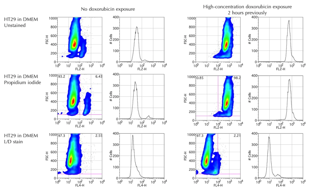
The most frequent error when assaying doxorubicin-treated samples is deploying conventional Annexin V/PI kits without accounting for the photophysical properties of the chemotherapeutic agent itself. Propidium iodide is a classical dye exclusion agent intended to enter only cells with compromised plasma membranes. However, in cells exposed to doxorubicin, high fluorescence intensity is registered in the PI collection channel even in viable, early apoptotic, or physically intact cells within hours of exposure [1]. Researchers subsequently misclassify viable drug-loaded cells as dead or necrotic.
Spectral Spillover into the Propidium Iodide Detector Channel
Doxorubicin has broad excitation and emission spectra [1, 2]. It is efficiently excited by the standard 488 nm blue laser typically used to excite both FITC and PI [1, 5]. Upon 488 nm excitation, doxorubicin exhibits wide fluorescent emission spanning from 530 nm to well beyond 650 nm, with an emission peak that overlaps almost completely with standard bandpass filters configured for PI (585/42 nm, FL2) and PE-Texas Red channels [1, 3].
As demonstrated by Lyon et al., HT29 cancer cells exposed to doxorubicin for just 2 hours display near 100% false-positive "death" in the FL2 channel because of this spectral overlap, despite the fact that 2 hours is biologically insufficient to execute necrotic membrane disruption [1]. Conversely, interrogating the identical cells via an independent far-red channel (FL4, 633/660 nm) reveals that the vast majority of cells remain physically intact and viable [1].
Rapid Intracellular Anthracycline Uptake and Nuclear Accumulation
The underlying physicochemical driver of this artifact is doxorubicin's planar tetracyclic anthraquinone chromophore structure. This hydrophobic core permits rapid passive membrane permeation and avid intercalation into cellular nucleic acids within minutes to hours of exposure [3, 4]. Because doxorubicin accumulates heavily inside viable cells, its intracellular concentration yields an intense endogenous fluorophore signal [4, 5]. When conventional flow cytometry compensation matrices are calculated solely using untreated control beads or untreated cells, the instrument cannot compensate for the massive, cell-associated doxorubicin emission across the 580–620 nm region, inevitably leading to false quadrant distribution and erroneous apoptosis calculations [1, 2].
How Can I Avoid This in Future Experiments?
Before running multi-color flow cytometry panels on compounds with chromophoric ring structures (such as anthracyclines, mitoxantrone, or curcuminoids), always run a single-cell spectral scan or an unstained drug-treated pilot tube to identify every channel impacted by drug autofluorescence. If an agent displays broad red-orange emission, proactively redesign your staining matrix by utilizing fluorophores detected outside the 550–620 nm window, prioritizing violet-excited (405 nm) or deep-red-excited (633–640 nm) reagents [1, 3].
Additionally, validate flow cytometry apoptosis measurements with orthogonal non-fluorescent or imaging-based assays—such as western blotting for cleaved PARP and cleaved caspase-3, or brightfield Hoechst morphological nuclear condensation scoring—to confirm true apoptotic commitment without optical interference [2, 3].
References
- [1]Lyon PC, et al. Scientific reports. 2021 Feb;11(1):4404.[IF: 4.996]
- [2]Wang Y, et al. Journal of experimental & clinical cancer research : CR. 2017 Aug;36(1):111.
- [3]Dhungel L, et al. Molecules (Basel, Switzerland). 2024 Feb;29(4).[IF: 4.6]
- [4]Perillo E, et al. Oncotarget. 2016 Jan;7(4):4077-92.[IF: 5.168]
- [5]Bognanni N, et al. Molecules (Basel, Switzerland). 2021 Oct;26(19).[IF: 4.927]