
Why did Doxorubicin-induced gamma-H2AX foci fail to appear in my Topo II-deficient cells?
Quick Answer
Recommended ApproachTo evaluate genotoxic stress in topoisomerase II-deficient cells, verify basal target expression and assess topoisomerase-independent readouts such as DNA adducts or oxidative lesions rather than canonical double-strand break markers.
Common CausesDiscrete gamma-H2AX foci failed to form because Doxorubicin requires the physical presence of topoisomerase II to trap covalent cleavable complexes that convert into double-strand breaks.
How Should It Be Done Correctly?
Experimental Parameters for Assessing Genotoxic Response
The following matrix outlines standard experimental conditions, target dependencies, and readout requirements for analyzing Doxorubicin-induced genotoxicity across proficient versus deficient cell models. [1, 2, 3]
| Parameter | Topoisomerase II-Proficient Model | Topoisomerase II-Deficient Model | Analytical Method / Specification |
|---|---|---|---|
| Target Abundance | High endogenous TOP2A / TOP2B expression | Knockdown, knockout, or low expression | Immunoblotting / RT-qPCR baseline validation [3] |
| Doxorubicin Concentration | 0.1–2.0 µM (cell line-dependent) | 0.1–2.0 µM (for direct comparison) | Working dilution prepared in pre-warmed media [1, 2, 3] |
| Vehicle / Stock Solution | 10 mM stock in anhydrous DMSO | 10 mM stock in anhydrous DMSO | Stored at -80°C (up to 1 year) or -20°C (1 month) |
| Primary Mechanism | TOP2 cleavable complex trapping | Topoisomerase-independent lesions | Covalent complex stabilization vs intercalation [3, 4] |
| Double-Strand Breaks (DSBs) | Robust, time-dependent formation | Minimal or absent frank DSBs | Neutral comet assay or pulsed-field electrophoresis [1, 5] |
| Primary DDR Readout | Discrete γ-H2AX foci (Ser139) | Minimal discrete γ-H2AX foci | Confocal immunofluorescence microscopy [1, 2, 6] |
| Alternative Readouts | Co-localized 53BP1 foci, cfDNA fragmentation | Direct DNA adducts, ROS, S-phase arrest | Flow cytometry, qPCR, or adduct assays [1, 3, 4] |
| Fixation / Permeabilization | 4% PFA (10 min) + 0.5% Triton X-100 | 4% PFA (10 min) + 0.5% Triton X-100 | Ice-cold PBS washes and standard blocking [2, 6] |
Validated Workflow for Detecting Topoisomerase-Mediated Damage
-
Verify Target Protein Expression Before Treatment Prior to applying anthracyclines, determine the precise protein levels of TOP2A and TOP2B in both wild-type control and target-deficient test lines via Western blot [3, 5]. If testing an engineered knockdown (e.g., siRNA or shRNA), quantify residual protein abundance to determine whether target suppression is partial or complete.
-
Reconstitution and Compound Preparation Prepare Doxorubicin stock solutions at 10 mM in anhydrous DMSO, ensuring complete dissolution. Store working aliquots tightly sealed at -80°C for up to 1 year or -20°C for up to 1 month. Immediately before exposure, dilute the stock directly into pre-warmed complete growth medium with vigorous mixing to prevent micro-precipitation.
-
Pulse Treatment and Kinetic Sampling Treat cells in parallel across identical time intervals (typically 1 to 8 hours for acute damage kinetics, or up to 24–48 hours for cell-cycle and senescence outcomes) [1, 2, 6]. Include a known topoisomerase-independent double-strand break control (such as bleomycin, ionizing radiation, or neocarzinostatin) alongside vehicle-treated controls (DMSO) to establish that your ATM/ATR cascade and histone H2AX phosphorylation machinery are functionally intact [1, 7].
-
Immunofluorescence Staining and Imaging Following treatment, wash cells gently with cold PBS and fix with 4% paraformaldehyde for 10 minutes at room temperature (or cold methanol at -20°C) [2, 6]. Permeabilize cell membranes using 0.5% Triton X-100 in PBS, followed by blocking with 1–2% BSA and normal goat serum [2, 6]. Incubate with a high-affinity anti-γ-H2AX (phospho-Ser139) primary antibody overnight at 4°C, followed by fluorophore-conjugated secondary antibody incubation and Hoechst or DAPI nuclear counterstaining [2, 6].
-
Quantitative Focus Analysis Acquire z-stack images on a confocal or automated high-content fluorescence microscope [1, 2]. Quantify both the percentage of focus-positive nuclei and the absolute focus count per individual cell rather than relying exclusively on total nuclear mean fluorescence intensity [1, 2].
What Went Wrong?
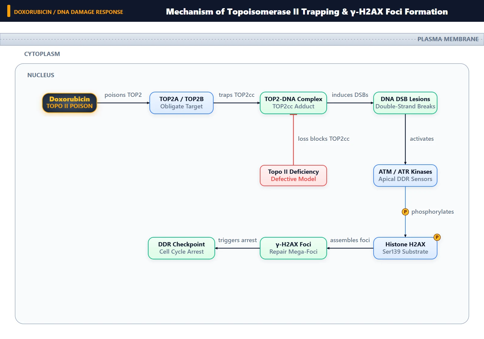
Copy code to add this image to your page Copy Code
Erroneous Expectation of Double-Strand Breaks Without Molecular Target
The fundamental mistake was assuming that Doxorubicin generates frank DNA double-strand breaks through a drug-autonomous, direct-cleavage mechanism. Doxorubicin is not a direct chemical endonuclease or radiomimetic agent; it relies on an endogenous protein target to cause double-strand cleavage [3, 5]. Expecting discrete γ-H2AX foci to appear in Topo II-depleted or knockout cells overlooks the direct causal chain connecting topoisomerase II engagement to DNA backbone rupture [3, 5, 8].
Failure of Cleavable Complex Trapping and Downstream Phosphorylation
Because the cellular abundance of topoisomerase II was depleted or abolished, Doxorubicin could not stabilize the covalent enzyme-DNA intermediate ("cleavable complex") [1, 3, 8]. In wild-type cells, these stabilized complexes stall transcription and replication machinery, causing permanent structural breaks that immediately trigger the activation of phosphoinositide 3-kinase-related protein kinases (principally ATM and ATR) [7, 8]. ATM and ATR directly phosphorylate histone H2AX at Ser139 across megabase chromatin domains surrounding each break [1, 7]. In the absence of Topo II, cleavable complexes cannot form, preventing ATM/ATR recruitment to local rupture sites and resulting in an absence of detectable γ-H2AX focus accumulation [1, 3, 7].
Topoisomerase II Dependence of Anthracycline-Induced Breakpoints
Doxorubicin operates via two distinct modes of action: topoisomerase II-dependent enzyme poisoning and topoisomerase II-independent physicochemical actions [3, 4, 5].
Under physiological and therapeutic concentrations, the primary cytotoxic pathway is the poisoning of topoisomerase II α and β [3, 4]. During normal catalytic cycles, Topo II creates a transient, reversible double-strand break with phosphotyrosyl bonds, passes an intact DNA duplex through the gate, and religates the backbone [8]. Doxorubicin intercalates at the scission site and traps the religation gate open [4, 8]. When replication forks or RNA polymerases collide with these stabilized complexes, irreparable double-strand breaks are generated, demarcated by γ-H2AX and 53BP1 foci [1, 3, 8].
In Topo II-deficient cells, Doxorubicin can still intercalate into parental DNA, generate reactive oxygen species (ROS), and produce covalent formaldehyde-mediated adducts, which stall replication forks in a Topo II-independent manner [3, 4, 5]. However, these lesions do not inherently create acute, frank double-strand breaks [3, 5]. Instead of generating sharp, distinct γ-H2AX focus clusters, they may produce only faint, diffuse pan-nuclear backgrounds or delayed single-strand lesions, explaining why discrete γ-H2AX foci failed to appear in your assay [1, 3, 5].
How Can I Avoid This in Future Experiments?
- Pre-screen Target Competence: Always quantify TOP2A and TOP2B expression by immunoblotting alongside any panel of cell lines before utilizing topoisomerase poisons, confirming whether the genetic background is permissive for cleavable complex formation [3, 5].
- Incorporate Target-Independent Positive Controls: Run parallel wells exposed to an agent that induces double-strand breaks independently of topoisomerases (such as bleomycin, calicheamicin, or ionizing radiation) to confirm that the cellular DNA damage response machinery and γ-H2AX staining workflow are operating correctly.
- Shift to Alternative Readouts in Deficient Backgrounds: When investigating Doxorubicin effects in Topo II-deficient cells, evaluate topoisomerase-independent endpoints—such as reactive oxygen species generation, alkaline comet assays for single-strand breaks, cell-cycle progression dynamics, or direct adduction assays—rather than canonical double-strand break markers [3, 4, 5].
References
- [1]Linke C, et al. Cancers. 2024 Nov;16(22).[IF: 4.4]
- [2]Strzeszewska-Potyrała A, et al. Cancers. 2021 Apr;13(9).[IF: 6.575]
- [3]Tentner AR, et al. Molecular systems biology. 2012 Jan;8:568.[IF: 11.34]
- [4]Dohadwala TK, et al. Discoveries (Craiova, Romania). 2025 Jan;13(3):e216.
- [5]Borella F, et al. Current oncology (Toronto, Ont.). 2024 Dec;31(12):8054-8074.[IF: 3.4]
- [6]Choi W, et al. Frontiers in oncology. 2025 Jan;15:1634503.[IF: 3.4]
- [7]Linders AN, et al. npj aging. 2024 Jan;10(1):9.[IF: 6.0]
- [8]Baguley BC, et al. Molecules (Basel, Switzerland). 2021 Jan;26(3).[IF: 4.927]