
Why does 5 µM Doxorubicin artificially increase background absorbance in my MTT viability assay?
Quick Answer
Recommended ApproachAspirate the Doxorubicin-containing culture medium and wash the cell monolayer thoroughly with PBS before adding MTT dissolved in PBS to eliminate residual colored drug.
Common CausesAdding MTT reagent directly to culture wells without washing allowed residual 5 µM Doxorubicin to persist, whose intrinsic chromophore absorbance spectrum directly overlaps with the formazan readout wavelength around 560–570 nm.
How Should It Be Done Correctly?
Optimization Parameters for Mitigating Doxorubicin Optical Interference
To eliminate optical crosstalk between Doxorubicin and formazan during viability testing, specific assay controls and washing modifications must be implemented [1].
| Assay Step / Parameter | Conventional MTT Protocol | Interference-Free Modified Protocol | Functional Purpose |
|---|---|---|---|
| Drug Removal Prior to MTT | No wash; reagent added directly to medium | Complete aspiration followed by 1–2× warm PBS wash | Eliminates extracellular 5 µM Doxorubicin prior to colorimetric development [1] |
| MTT Incubation Matrix | Complete culture medium (e.g., DMEM with phenol red) | Clear balanced salt solution (sterile PBS or phenol red-free medium) | Prevents additive absorbance from medium components at ~560 nm [1] |
| Drug-Only Control Wells | Usually omitted | Parallel cell-free wells containing 5 µM Doxorubicin in vehicle | Establishes drug-specific baseline optical density to subtract if needed |
| Measurement Wavelength | Single wavelength (560–570 nm) | Dual wavelength: 560–570 nm (signal) minus 650–690 nm (reference) | Corrects for non-specific optical scatter, plate imperfections, and fingerprints [2, 3] |
| Solubilization Check | Visual confirmation of dissolution | Microplate agitation (15–30 min) in DMSO or acidified isopropanol | Ensures complete dissolution of formazan crystals before reading [1, 3] |
Step-by-Step Interference-Free MTT Protocol
-
Treatment Termination and Aspiration At the end of the Doxorubicin exposure window (e.g., 24 h or 48 h), carefully remove the multi-well plate from the incubator [1, 2]. Using an aspirator or multichannel pipette, remove the conditioned medium containing 5 µM Doxorubicin completely from each well, taking care not to touch or scratch the adherent cell layer.
-
Gentle PBS Wash Step Gently add 100–200 µL of sterile, pre-warmed (37°C) 1× PBS to each well. Swirl the plate gently for 30 seconds and aspirate completely. Repeating this wash once more ensures the total removal of residual unbound drug, which otherwise persists on the plastic surface or meniscus and interferes with absorption [1].
-
Application of MTT Reagent in Buffer Reconstitute MTT stock solution in sterile PBS at 5 mg/mL, sterile filter (0.22 µm), and dilute it into pre-warmed PBS (or phenol red-free basal medium) to a working concentration of 0.5 mg/mL [1]. Dispense 100 µL into each well. Avoid serum-containing or phenol red-containing medium during this incubation to maintain a clear baseline [1].
-
Formazan Crystal Development Incubate the plates at 37°C in a 5% CO₂ humidified incubator for 2 to 4 hours. Inspect the wells under an inverted brightfield microscope: viable metabolically active cells will exhibit intracellular dark-purple needle-like or punctate formazan deposits, while dead cells will show cellular debris and lack crystal formation [3].
-
Formazan Solubilization and Dual-Wavelength Measurement Carefully aspirate the MTT solution without dislodging the crystal precipitates at the bottom of the wells. Add 100 µL of pure DMSO (or acidified isopropanol) to each well [1, 3]. Agitate the microplate on an orbital shaker at room temperature for 15–20 minutes protected from light. Read the absorbance on a microplate spectrophotometer at a primary measurement wavelength of 560–570 nm, paired with a reference wavelength of 650–690 nm [1, 2].
What Went Wrong?
Copy code to add this image to your page Copy Code
Direct Addition of MTT to Doxorubicin-Conditioned Medium
The fundamental procedural error was adding the tetrazolium dye directly into the culture wells without removing the 5 µM Doxorubicin treatment medium and without washing the cell layer [1]. In standard protocols designed for colorless xenobiotics, investigators frequently add concentrated MTT stock solution directly to the existing medium. However, when working with intrinsically chromophoric molecules at micromolar concentrations, residual drug solution remains present in the well volume or clings to the microplate walls during crystal solubilization [1].
Proximate Cause of the Falsely Elevated Absorbance Signal
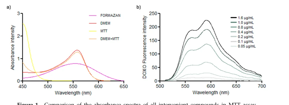
The artificial elevation in background absorbance occurs because Doxorubicin exhibits broad intrinsic absorption in the visible spectrum that directly overlaps with the absorption peak of dissolved purple formazan [1]. Because formazan is quantified spectrophotometrically around 560–570 nm, any unremoved Doxorubicin contributes directly to the measured optical density (OD) [1, 2]. At a concentration of 5 µM (equivalent to ~2.9 µg/mL), Doxorubicin yields significant absorbance at 560 nm. This optical contribution is erroneously recorded by the plate reader as metabolic conversion of MTT, manifesting as falsely elevated cell viability or distorted sigmoidal kill curves [1].
Underlying Physicochemical and Photometric Mechanisms
Chemically, Doxorubicin contains a conjugated planar anthraquinone chromophore system linked to an aminoglycoside moiety [4, 5]. This extensive conjugated tetracyclic quinone-hydroquinone structure gives Doxorubicin its characteristic deep red/orange pigmentation and confers visible light absorption across the 450–600 nm range, featuring significant absorbance and fluorescence emission overlapping at 560 nm [1].
When mitochondrial and cytosolic oxidoreductases in viable cells reduce the yellow tetrazolium salt (MTT) into insoluble purple formazan, the resulting crystals have an absorbance maximum centered precisely between 560 nm and 570 nm [1, 2]. When the final solubilization solvent (such as DMSO) is added, it dissolves both the intracellular formazan crystals and any remaining extracellular or cell-associated Doxorubicin [1]. Because absorbance is strictly additive according to the Beer-Lambert law:
$Aₜₒₜₐₗ = Afₒᵣₘₐzₐₙ + ADₒₓₒᵣᵤbᵢcᵢₙ + Aₘₑdᵢᵤₘ bₐcₖgᵣₒᵤₙd$
the spectrophotometer registers the sum of both chromophores [1]. At 5 µM, the drug's extinction coefficient at 560 nm is high enough to elevate baseline optical density dramatically, entirely masking the reduction in cell viability caused by Doxorubicin's cytotoxic actions [1, 4].
How Can I Avoid This in Future Experiments?
To ensure reliable cytotoxicity profiling with anthracyclines and other colored therapeutics, include drug-only background wells on every plate containing serial dilutions of Doxorubicin in medium without cells, treated identically to test wells. Subtracting these drug-specific blank OD values mathematically compensates for any residual compound binding to the plastic surface.
Alternatively, consider substituting the MTT assay with orthogonal viability methodologies that do not rely on absorbance in the 500–600 nm range. Bioluminescent ATP assays (e.g., CellTiter-Glo) quantify luciferase-catalyzed luminescence rather than optical density, making them completely immune to chromophoric and fluorescent spectral overlaps caused by Doxorubicin [6].
References
- [1]Luis C, et al. Methods and protocols. 2019 Apr;2(2).[IF: 2.4]
- [2]Druzhkova I, et al. Cancers. 2022 Nov;14(22).[IF: 5.2]
- [3]Liu T, et al. Oncotarget. 2016 Dec;7(50):83502-83513.[IF: 5.168]
- [4]Kciuk M, et al. Cells. 2023 Feb;12(4).[IF: 5.1]
- [5]Peter S, et al. Molecules (Basel, Switzerland). 2022 Jul;27(14).[IF: 4.6]
- [6]Dilz J, et al. Scientific reports. 2023 Apr;13(1):6167.[IF: 3.8]