
Why did prolonged light exposure cause my red Doxorubicin working solution to fade and lose potency?
Quick Answer
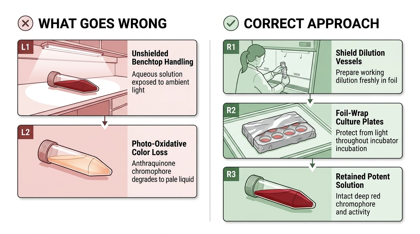
Recommended ApproachPrepare doxorubicin working solutions immediately before use from light-shielded DMSO stocks, strictly handling and incubating all dilutions inside foil-wrapped or amber vessels.
Common CausesProlonged exposure of the working solution to ambient benchtop light caused photo-oxidative breakdown of doxorubicin's red anthraquinone chromophore into inactive degradation products.
How Should It Be Done Correctly?
Copy code to add this image to your page Copy Code
Handling Parameters and Storage Specifications
Official storage conditions, preparation guidelines, and photoprotection controls for doxorubicin stock and working solutions.
Step-by-Step Preparation and Photoprotective Protocol
- Reconstitute concentrated stock in a darkened hood: Weigh lyophilized powder under subdued room lighting and dissolve in high-grade anhydrous DMSO to achieve the manufacturer-recommended stock concentration. Invert or gently vortex until the compound fully dissolves into a clear, deep red-orange solution without particulates.
- Aliquot into light-impermeable vessels: Immediately partition the freshly dissolved stock into small single-use aliquots using pre-chilled amber microcentrifuge tubes or standard low-binding tubes thoroughly wrapped in heavy-duty aluminum foil. Purge the headspace with dry nitrogen or argon before sealing to minimize dissolved oxygen, and immediately transfer aliquots to -80°C.
-
Prepare working solutions immediately before use: Thaw a single stock aliquot rapidly at room temperature protected from light. Dilute into pre-warmed culture medium or balanced salt solution immediately prior to cell treatment; never prepare aqueous working dilutions ahead of time or store them overnight at 4°C, as diluted aqueous doxorubicin exhibits marked instability compared to frozen neat DMSO stocks [1].
-
Maintain strict photoprotection during administration: Shield all dilution tubes, pipettes, and multi-channel reservoirs from direct biosafety cabinet lighting by turning off cabinet fluorescent/UV lamps or shading vessels with aluminum foil hoods. When administering to culture vessels, work quickly under dimmed ambient lighting.
- Shield cell culture vessels during incubation: Wrap tissue culture plates or culture flasks in aluminum foil prior to placing them into the 37°C humidified incubator. Maintain light protection across all post-treatment washing, imaging, or harvesting steps until cell lysis or fixation is complete.
| Operational Parameter | Specification / Standard Setting | Container & Handling Environment | Stability & Usable Window |
|---|---|---|---|
| Solid Powder Storage | -20°C, desiccated | Original amber vial or light-proof desiccator | 3 years |
| Stock Reconstitution | 10 mM in DMSO (5.7998 mg/mL) | Anhydrous DMSO, sterile amber tubes | Single-use working aliquots |
| Stock Solution Storage | -80°C (preferred) or -20°C | Hermetically sealed, aluminum foil wrap | 1 year at -80°C; 1 month at -20°C |
| Working Dilution Media | Aqueous buffer / culture medium (pH 6.5–7.4) | Clear or amber tubes wrapped in aluminum foil | Prepare freshly (<4 h working window) |
| Light Shielding Controls | Complete exclusion of ambient visible/UV light | Foil wrap, amber plastics, dimmed biosafety cabinet | Continuous throughout handling and treatment |
What Went Wrong?
Unshielded Light Exposure on the Open Benchtop
The red working solution was left in transparent plastic or glass vessels exposed to ambient laboratory lighting or direct sunlight over an extended period. Because aqueous dilutions of doxorubicin are far more vulnerable to ambient conditions than frozen organic stocks, keeping an unprotected low-concentration solution on the open benchtop accelerated degradation within hours [1].
Photo-Induced Decomposition and Color Fading
The visible fading from intense red to pale orange or colorless liquid is the direct phenotypic indicator that the core conjugated system has been destroyed. In aqueous solutions, exposure to indirect sunlight for 10 hours degrades doxorubicin down to 62.4% of its starting concentration, progressing to comprehensive breakdown upon longer light exposure [1]. The loss of cytotoxic potency directly tracks this structural decomposition: once the molecule breaks down, it loses its ability to engage target DNA and topoisomerase II complexes [1, 2].
Photo-Oxidative Ring Cleavage and Radical Generation
Doxorubicin's characteristic dark red pigment originates from its planar tetracyclic anthraquinone chromophore [2, 3]. Anthraquinones are efficient photoactive sensitizers: upon absorbing visible and near-ultraviolet photons (broad absorption bands around 480–500 nm), the conjugated tetracenequinone core transitions to an excited triplet state [1, 3, 4].
This photo-excited species reacts directly with dissolved molecular oxygen and surrounding water molecules via redox cycling, generating reactive oxygen species (ROS) such as singlet oxygen, superoxide radicals, and hydrogen peroxide [2, 3, 4]. The localized generation of peroxides triggers oxidative hydroxylation of the phenolic benzene rings at ortho- and para-positions relative to the methoxy substituent [1]. This oxidative cleavage dismantles the conjugated polycyclic electron system, destroying the visible chromophore and detaching the daunosamine aminosugar side chain [1, 4]. Because intercalation into DNA base pairs and stabilization of the topoisomerase II cleavage complex require both the intact planar tetracyclic ring system and the basic daunosamine sugar [2, 3, 4], photo-fragmented products are pharmacologically inactive.
How Can I Avoid This in Future Experiments?
- Adopt dedicated amber consumables: Standardize all working steps on amber polypropylene microcentrifuge tubes and black-walled or foil-wrapped multi-well culture plates to eliminate human error associated with loose foil wraps.
- Enforce a strict single-use dilution policy: Treat doxorubicin working solutions strictly as single-experiment reagents; calculate the exact volume required for the day's dosing schedule and discard any unused aqueous residue rather than saving it for subsequent treatment timepoints.
- Incorporate an optical density check: Verify the integrity of questionable working solutions by measuring their absorbance profile; intact doxorubicin exhibits distinct absorbance maxima around 480–495 nm, and any flattening or bluing/hypochromic shift indicates unacceptable photo-oxidative degradation [1, 3].