
Why do my MDR1-overexpressing cancer cells show no nuclear accumulation of 1 µM Doxorubicin?
Quick Answer
Recommended ApproachPre-treat MDR1-overexpressing cells with an ABCB1 inhibitor such as 10 µM verapamil or evaluate parental control cells in parallel to block transporter-mediated efflux and restore nuclear doxorubicin accumulation.
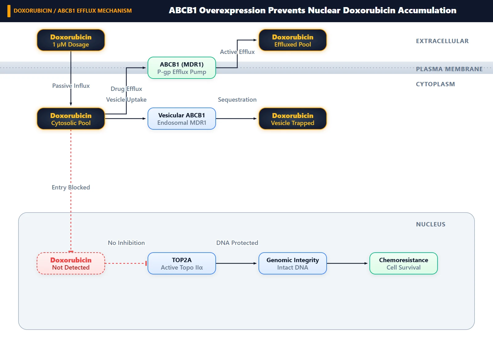
Common CausesOverexpressed ABCB1 actively pumps doxorubicin out of the cell and sequesters intracellular drug within cytoplasmic vesicles, entirely preventing nuclear entry and DNA intercalation.
How Should It Be Done Correctly?
Quantitative Parameters for Transporter and Accumulation Assays
To evaluate doxorubicin uptake and verify whether poor nuclear accumulation stems from multidrug resistance protein 1 (MDR1/ABCB1) activity, the assay must be conducted with appropriate drug concentrations, validated transporter inhibitors, and proper nuclear counterstaining.
| Parameter | Baseline Condition | Transporter Inhibition Control | Operational Specifications / Notes |
|---|---|---|---|
| Stock Reagent | 10 mM in DMSO | N/A | Dissolve powder (MW 579.98) at 5.7998 mg/mL in DMSO; store stock at -80°C (1 year) or -20°C (1 month) |
| Doxorubicin Dosing | 1 µM working solution | 1 µM working solution | Dilute stock freshly into pre-warmed culture medium; keep vehicle DMSO ≤0.1% (v/v) [1, 2] |
| Efflux Inhibitor | Vehicle (DMSO / PBS) | Verapamil (10 µM) or Adagrasib (1 µM) | Pre-incubate 1–2 h prior to doxorubicin exposure to fully occupy ABCB1 substrate-binding sites [3, 4] |
| Treatment Duration | 2 h to 24 h | 2 h to 24 h | Short exposure (2–4 h) measures acute uptake; extended exposure (24 h) measures steady-state compartmentalization [1, 4] |
| Nuclear Counterstain | DRAQ5 (5 µM) or DAPI (1 µg/mL) | DRAQ5 (5 µM) or DAPI (1 µg/mL) | Counterstain for 15 min at 37°C or ambient temperature to demarcate the nuclear compartment [5] |
| Optical Readout | Ex 488 nm / Em 575–595 nm | Ex 488 nm / Em 575–595 nm | Detect native doxorubicin anthracycline fluorescence via confocal microscopy or flow cytometry [1, 3, 4] |
Step-by-Step Procedure for Visualizing Nuclear Drug Penetration
- Seed Experimental and Control Cells: Plate both parental (drug-sensitive or empty-vector control) and MDR1-overexpressing cells at 60–70% confluency in glass-bottom confocal dishes or multi-well plates [1, 3]. Allow cells to adhere overnight under standard growth conditions.
- Apply Transporter Inhibitor: Replace the growth medium in designated control wells with medium containing 10 µM verapamil or 1 µM adagrasib (MRTX849) [3, 4]. Incubate for 1 h at 37°C before introducing doxorubicin to achieve complete transporter saturation.
- Administer Doxorubicin: Add doxorubicin to all test and control wells at a final concentration of 1 µM, ensuring thorough mixing by gentle plate swirling [1, 2]. Protect the culture vessels from direct light during incubation.
- Maintain Light-Protected Incubation: Incubate the cells for 2 to 24 h at 37°C. For acute efflux kinetics, shorter timepoints (2–4 h) distinguish initial transport from secondary metabolic conversion [5, 6].
- Counterstain and Wash: Aspirate the drug-containing medium and immediately rinse the cell layer twice with ice-cold PBS to halt further transport. Apply DRAQ5 or DAPI for 15 min according to standard labeling protocols to define the nuclear boundaries [5].
- Confocal Imaging and Quantification: Image living or fixed cells using a laser scanning confocal microscope (Ex 488 nm, Em 590 nm for doxorubicin; Ex 633 nm for DRAQ5) [4, 5]. Quantify the ratio of nuclear to cytoplasmic mean fluorescence intensity across multiple randomly selected fields of view.
What Went Wrong?
Copy code to add this image to your page Copy Code
Presumption of Unhindered Nuclear Translocation
The failure to detect doxorubicin in the nucleus of MDR1-overexpressing cells stems from the incorrect assumption that 1 µM doxorubicin can passively diffuse into the nucleoplasm irrespective of ABC transporter expression. Testing 1 µM doxorubicin as a single agent without including an ABCB1 inhibitor (such as verapamil) or a drug-sensitive parental control prevents discrimination between failed cellular entry, accelerated efflux, and organelle entrapment [3, 4]. Researchers frequently expect bright nuclear signal because wild-type cells readily display nuclear fluorescence at 1 µM, overlooking that overexpressed ABCB1 completely alters anthracycline pharmacokinetics [1, 4].
Active Transporter Efflux and Cytoplasmic Sequestration
The proximate reason doxorubicin fails to reach the nucleus is twofold: robust extrusion across the plasma membrane and spatial redirection away from the nuclear envelope [3, 4]. In cells overexpressing MDR1 (ABCB1/P-glycoprotein), the rate of ATP-dependent drug efflux exceeds passive membrane influx, dramatically lowering total intracellular drug accumulation [1, 3]. Furthermore, whatever small fraction of doxorubicin manages to enter the cytoplasm is intercepted and pumped out of the nucleoplasm into perinuclear vesicular structures, endosomes, and lysosomes, resulting in faint cytoplasmic puncta rather than diffuse nuclear staining [4, 5].
Transporter-Driven Target Shielding and Energetics
Doxorubicin is a hydrophobic, amphiphilic, positively charged anthracycline that acts as an optimal substrate for the ABCB1 efflux pump [2]. ABCB1 utilizes energy derived from ATP binding and hydrolysis at its nucleotide-binding domains (NBDs) to export doxorubicin through its transmembrane domains into the extracellular space [3, 6].
In addition to plasma membrane extrusion, ABCB1 localized on intracellular membranes and perinuclear organelles actively pumps doxorubicin into vesicular lumens, preventing the drug from intercalating into genomic DNA or reaching topoisomerase II (TOP2A) [4, 5, 7]. Furthermore, drug-resistant cancer lines often show altered carbonyl reduction pathways that convert doxorubicin into doxorubicinol, a metabolite with markedly lower DNA-binding affinity that remains trapped in extranuclear compartments [5, 7]. Consequently, the nuclear compartment remains completely shielded unless ABCB1 pump activity is disrupted [3, 4].
How Can I Improve the Experiment?
Always incorporate a parallel drug-sensitive parental line (e.g., empty-vector control) alongside the MDR1-overexpressing line. Comparing both lines treated with 1 µM doxorubicin under identical acquisition settings immediately confirms whether the absence of nuclear signal is an MDR1-dependent phenotype [1, 3].
Pair confocal imaging with live-cell organelle trackers (such as DiO membrane dye or LysoTracker Green) to confirm whether doxorubicin fluorescence colocalizes with cytoplasmic vesicles or lysosomes [4, 5]. If vesicular sequestration is prominent, assessing doxorubicin efflux kinetics by flow cytometry provides quantitative, high-throughput confirmation of transporter activity across thousands of cells [1, 3].
References
- [1]Tomar MS, et al. Frontiers in pharmacology. 2026 Jan;17:1760920.[IF: 5.4]
- [2]Arnason T, et al. Cancers. 2015 Oct;7(4):2063-82.[IF: 5.326]
- [3]Zhang Y, et al. Cell communication and signaling : CCS. 2022 Sep;20(1):142.[IF: 8.4]
- [4]Barbieri F, et al. BMC cancer. 2015 Apr;15:228.[IF: 3.265]
- [5]Heibein AD, et al. BMC cancer. 2012 Aug;12:381.[IF: 3.333]
- [6]Steelman LS, et al. Oncotarget. 2017 Dec;8(68):113013-113033.[IF: 5.168]
- [7]Piska K, et al. Investigational new drugs. 2017 Jun;35(3):375-385.[IF: 3.502]