-
 Australia
-
 Austria
-
 Belgium
-
 Brazil
-
 Canada
-
 China
China
-
 Czech Republic
-
 Denmark
-
 Finland
-
 France
-
 Germany
-
 Greece
-
 Hong Kong
-
 Hungary
-
 Iceland
-
 India
India
-
 Ireland
-
 Israel
-
 Italy
Italy
-
 Japan
Japan
-
 Korea
Korea
-
 Luxembourg
-
 Malaysia
-
 Netherlands
-
 New Zealand
-
 Norway
-
 Poland
-
 Qatar
Qatar
-
 Romania
-
 Saudi Arabia
-
 Singapore
-
 Spain
Spain
-
 Sweden
-
 Switzerland
-
 Taiwan
-
 Turkey
-
 United Kingdom
-
 United States
research use only
Chaetocin Histone Methyltransferase inhibitor
Cat.No.S8068
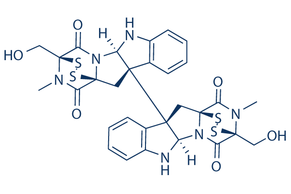
Chemical Structure
Molecular Weight: 696.84
Quality Control
| Related Targets | HDAC JAK BET PKC PARP HIF PRMT EZH2 AMPK Histone Acetyltransferase |
|---|---|
| Other Histone Methyltransferase Inhibitors | Pinometostat (EPZ5676) 3-Deazaneplanocin A (DZNep) Hydrochloride BIX-01294 Trihydrochloride EPZ015666 (GSK3235025) UNC1999 EPZ004777 MM-102 (HMTase Inhibitor IX) SGC 0946 EPZ005687 UNC0638 |
Cell Culture, Treatment & Working Concentration
| Cell Lines | Assay Type | Concentration | Incubation Time | Formulation | Activity Description | PMID |
|---|---|---|---|---|---|---|
| A549 | Cytotoxicity assay | Cytotoxicity against human A549 cells, IC50 = 0.025 μM. | 20303767 | |||
| Hep3b | Function assay | Inhibition of HIF-1alpha-mediated VEGF expression in human Hep3b cells by luciferase reporter gene assay, IC50 = 0.04 μM. | 22305612 | |||
| Hep3b | Function assay | Inhibition of HIF-1alpha-mediated VEGF secretion in human Hep3b cells by ELISA, IC50 = 0.1 μM. | 22305612 | |||
| Jurkat | Cytotoxicity assay | 40 hrs | Cytotoxicity against human Jurkat cells after 40 hrs bioluminescence assay, IC50 = 0.6 μM. | 20303767 | ||
| Drosophila melanogaster SL2 | Function assay | 0.1 uM | 5 days | Inhibition of SU(VAR)3-9 in Drosophila melanogaster SL2 cells assessed as reduction of H3K9me2 at 0.1 uM after 5 days by MALDI-TOF MS seeded at 2.5 million cells/ml density | 16408017 | |
| Drosophila melanogaster SL2 | Function assay | 0.5 uM | 5 days | Inhibition of SU(VAR)3-9 in Drosophila melanogaster SL2 cells assessed as reduction of H3K9me2 at 0.5 uM after 5 days by MALDI-TOF MS seeded at 2.5 million cells/ml density | 16408017 | |
| Drosophila melanogaster SL2 | Function assay | 0.5 uM | Inhibition of SU(VAR)3-9 in Drosophila melanogaster SL2 cells assessed as reduction of H3K9me3 at 0.5 uM by immunostaining method | 16408017 | ||
| HL60 | Apoptosis assay | 30 uM | 4 hrs | Induction of apoptosis in human HL60 cells assessed as necrotic cell death at 30 uM after 4 hrs using DAPI/PI by confocal laser microscopy analysis | 20675131 | |
| HL60 | Apoptosis assay | 0.3 uM | 4 hrs | Induction of apoptosis in human HL60 cells assessed as nuclear fragmentation at 0.3 uM after 4 hrs using DAPI/PI by confocal laser microscopy analysis | 20675131 | |
| HL60 | Apoptosis assay | 0.3 uM | 2 hrs | Induction of apoptosis in human HL60 cells assessed as activation of caspase 3 at 0.3 uM after 2 hrs by Western blot analysis | 20675131 | |
| MCF7 | Function assay | Inhibition of histone-lysine N-methyltransferase in human MCF7 cells assessed as decrease in methylation of RARbeta DNA by qPCR method | ChEMBL | |||
| Click to View More Cell Line Experimental Data | ||||||
Solubility
|
In vitro |
DMSO
: 25 mg/mL
(35.87 mM)
Water : Insoluble Ethanol : Insoluble |
Molarity Calculator
|
In vivo |
|||||
In vivo Formulation Calculator (Clear solution)
Step 1: Enter information below (Recommended: An additional animal making an allowance for loss during the experiment)
Step 2: Enter the in vivo formulation (This is only the calculator, not formulation. Please contact us first if there is no in vivo formulation at the solubility Section.)
Calculation results:
Working concentration: mg/ml;
Method for preparing DMSO master liquid: mg drug pre-dissolved in μL DMSO ( Master liquid concentration mg/mL, Please contact us first if the concentration exceeds the DMSO solubility of the batch of drug. )
Method for preparing in vivo formulation: Take μL DMSO master liquid, next addμL PEG300, mix and clarify, next addμL Tween 80, mix and clarify, next add μL ddH2O, mix and clarify.
Method for preparing in vivo formulation: Take μL DMSO master liquid, next add μL Corn oil, mix and clarify.
Note: 1. Please make sure the liquid is clear before adding the next solvent.
2. Be sure to add the solvent(s) in order. You must ensure that the solution obtained, in the previous addition, is a clear solution before proceeding to add the next solvent. Physical methods such
as vortex, ultrasound or hot water bath can be used to aid dissolving.
Chemical Information, Storage & Stability
| Molecular Weight | 696.84 | Formula | C30H28N6O6S4 |
Storage (From the date of receipt) | |
|---|---|---|---|---|---|
| CAS No. | 28097-03-2 | Download SDF | Storage of Stock Solutions |
|
|
Mechanism of Action
| Targets/IC50/Ki |
dSU(VAR)3-9
0.8 μM
mouse G9a
2.5 μM
Neurospora crassa DIM5
3 μM
|
|---|---|
| In vitro |
In SL-2 Drosophila tissue culture cells, Chaetocin causes the inhibition of SU(VAR)3-9 and the number of H3 molecules dimethylated at Lys9, and also inhibits cell growth. In HepG2, Hep3B, and Huh7 human hepatoma cells, this compound inhibits HIF-1-Mediated hypoxic responses. It also potently inhibits proliferation and colony formation in a broad range of cancer cell lines with IC50 of 2-10 nM. |
| Kinase Assay |
Methylation assays
|
|
Unless otherwise stated, reactions are performed as followed: 1 g of purified enzyme (dSU(VAR)3-9 213; GST-hSUV39H1(82-412); GST-ncDIM5(19-318) and GSTmG9a(621-1000); dPRSET7(1-691); His-SET7/9(109-366) or enzyme complex expressed in a baculoviral expression system (dE(z), dSU(Z)12, dp55, dESC) were incubated for 30 minutes with 1 g of a peptide containing the first 19 amino acids of H3 plus an additional cystein residue (ARTKQTARKSTGGKAPRKQC) in a total volume of 40μl in BC25 (10mM HEPES pH7.6, 25mM NaCl, 1mM EDTA, 10%glycerol). All enzymes except for the E(z) complex had a similar specific activity between 5-10 nmole/min/mg. For the E(z) complex, the specific activity is approximately 5-10 fold lower, which is probably due to an incomplete formation of the full complex that is required for the highest activity. As a methyl donor S-Adenosyl [methyl-3H] methionine (SAM) is present in the reaction at a final concentration of 40 M with a specific activity of 0.3Ci/mmole. Reactions are stopped by adding 1/10 volume of 100% acetic acid and the incorporation of radioactivity is measured by spotting 30 l of the reaction on P81 filter paper and subsequent scintillation counting. For the inhibitor screening, 1 l of this compound with a concentration of 10 g/l is added to each reaction. In the cases where PRSET7 or the E(z) complex is used as enzyme either recombinant nucleosomes (1 g) or recombinant H3 (1 g) respectively replaced the H3 peptide as a substrate.
|
|
| In vivo |
In Hepa 1c1c-7 tumor-bearing mice, Chaetocin (0.25 mg/kg, i.p.) inhibits tumor growth by deregulating HIF-1[alpha]-mediated angiogenesis. In SKOV3 tumor-bearing nude mice, this compound treatment (0.25 mg/kg, i.p.) significantly delays the tumor growth with minimal evidence of toxicities. |
References |
|
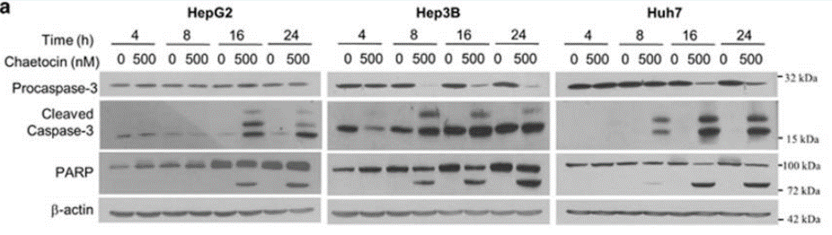
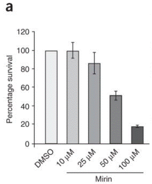
Applications
| Methods | Biomarkers | Images | PMID |
|---|---|---|---|
| Western blot | Procaspase-3 / Cleaved Caspase-3 / PARP |
|
26890137 |
| Growth inhibition assay | Cell viability |
|
18176557 |
Tech Support
Tel: +1-832-582-8158 Ext:3
If you have any other enquiries, please leave a message.
Signaling Pathway Map
Products are for research use only. Not for human use. We do not sell to patients.
©Copyright 2013 Selleck Chemicals. All Rights Reserved.