- Bioactive Compounds
- By Signaling Pathways
- PI3K/Akt/mTOR
- Epigenetics
- Methylation
- Immunology & Inflammation
- Protein Tyrosine Kinase
- Angiogenesis
- Apoptosis
- Autophagy
- ER stress & UPR
- JAK/STAT
- MAPK
- Cytoskeletal Signaling
- Cell Cycle
- TGF-beta/Smad
- Compound Libraries
- Popular Compound Libraries
- Customize Library
- Clinical and FDA-approved Related
- Bioactive Compound Libraries
- Inhibitor Related
- Natural Product Related
- Metabolism Related
- Cell Death Related
- By Signaling Pathway
- By Disease
- Anti-infection and Antiviral Related
- Neuronal and Immunology Related
- Fragment and Covalent Related
- FDA-approved Drug Library
- FDA-approved & Passed Phase I Drug Library
- Preclinical/Clinical Compound Library
- Bioactive Compound Library-I
- Bioactive Compound Library-Ⅱ
- Kinase Inhibitor Library
- Express-Pick Library
- Natural Product Library
- Human Endogenous Metabolite Compound Library
- Alkaloid Compound LibraryNew
- Angiogenesis Related compound Library
- Anti-Aging Compound Library
- Anti-alzheimer Disease Compound Library
- Antibiotics compound Library
- Anti-cancer Compound Library
- Anti-cancer Compound Library-Ⅱ
- Anti-cancer Metabolism Compound Library
- Anti-Cardiovascular Disease Compound Library
- Anti-diabetic Compound Library
- Anti-infection Compound Library
- Antioxidant Compound Library
- Anti-parasitic Compound Library
- Antiviral Compound Library
- Apoptosis Compound Library
- Autophagy Compound Library
- Calcium Channel Blocker LibraryNew
- Cambridge Cancer Compound Library
- Carbohydrate Metabolism Compound LibraryNew
- Cell Cycle compound library
- CNS-Penetrant Compound Library
- Covalent Inhibitor Library
- Cytokine Inhibitor LibraryNew
- Cytoskeletal Signaling Pathway Compound Library
- DNA Damage/DNA Repair compound Library
- Drug-like Compound Library
- Endoplasmic Reticulum Stress Compound Library
- Epigenetics Compound Library
- Exosome Secretion Related Compound LibraryNew
- FDA-approved Anticancer Drug LibraryNew
- Ferroptosis Compound Library
- Flavonoid Compound Library
- Fragment Library
- Glutamine Metabolism Compound Library
- Glycolysis Compound Library
- GPCR Compound Library
- Gut Microbial Metabolite Library
- HIF-1 Signaling Pathway Compound Library
- Highly Selective Inhibitor Library
- Histone modification compound library
- HTS Library for Drug Discovery
- Human Hormone Related Compound LibraryNew
- Human Transcription Factor Compound LibraryNew
- Immunology/Inflammation Compound Library
- Inhibitor Library
- Ion Channel Ligand Library
- JAK/STAT compound library
- Lipid Metabolism Compound LibraryNew
- Macrocyclic Compound Library
- MAPK Inhibitor Library
- Medicine Food Homology Compound Library
- Metabolism Compound Library
- Methylation Compound Library
- Mouse Metabolite Compound LibraryNew
- Natural Organic Compound Library
- Neuronal Signaling Compound Library
- NF-κB Signaling Compound Library
- Nucleoside Analogue Library
- Obesity Compound Library
- Oxidative Stress Compound LibraryNew
- Plant Extract Library
- Phenotypic Screening Library
- PI3K/Akt Inhibitor Library
- Protease Inhibitor Library
- Protein-protein Interaction Inhibitor Library
- Pyroptosis Compound Library
- Small Molecule Immuno-Oncology Compound Library
- Mitochondria-Targeted Compound LibraryNew
- Stem Cell Differentiation Compound LibraryNew
- Stem Cell Signaling Compound Library
- Natural Phenol Compound LibraryNew
- Natural Terpenoid Compound LibraryNew
- TGF-beta/Smad compound library
- Traditional Chinese Medicine Library
- Tyrosine Kinase Inhibitor Library
- Ubiquitination Compound Library
-
Cherry Picking
You can personalize your library with chemicals from within Selleck's inventory. Build the right library for your research endeavors by choosing from compounds in all of our available libraries.
Please contact us at [email protected] to customize your library.
You could select:
- Antibodies
- Bioreagents
- qPCR
- 2x SYBR Green qPCR Master Mix
- 2x SYBR Green qPCR Master Mix(Low ROX)
- 2x SYBR Green qPCR Master Mix(High ROX)
- Protein Assay
- Protein A/G Magnetic Beads for IP
- Anti-Flag magnetic beads
- Anti-Flag Affinity Gel
- Anti-Myc magnetic beads
- Anti-HA magnetic beads
- Poly FLAG Peptide lyophilized powder
- Protease Inhibitor Cocktail
- Protease Inhibitor Cocktail (EDTA-Free, 100X in DMSO)
- Phosphatase Inhibitor Cocktail (2 Tubes, 100X)
- Cell Biology
- Cell Counting Kit-8 (CCK-8)
- Animal Experiment
- Mouse Direct PCR Kit (For Genotyping)
- New Products
- Contact Us
Ulixertinib (BVD-523)
Synonyms: VRT752271
Ulixertinib (BVD-523, VRT752271) is a potent and reversible ERK1/ERK2 inhibitor with IC50 of <0.3 nM for ERK2. Phase 1.

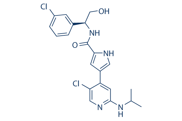
Ulixertinib (BVD-523) Chemical Structure
CAS: 869886-67-9
Selleck's Ulixertinib (BVD-523) has been cited by 56 publications
Purity & Quality Control
Batch:
Purity:
99.99%
99.99
Products often used together with Ulixertinib (BVD-523)
Ulixertinib, Encorafenib, and Cetuximab combo inhibits tumor growth better in CRC with BRAFV600E mutations.
Knoerzer D, et al. Cancer Res (2023) 83 (7_Supplement): 2693.
Ulixertinib and SHP099 inhibit downstream MAPK signaling in SK-N-AS and CHP-212 cells and result in tumor growth delay in xenograft mice model.
Valencia-Sama I, et al. Cancer Res. 2020 Aug 15;80(16):3413-3423.
Ulixertinib and Trametinib plus Trastuzumab deruxtecan are extremely effective and lead to complete tumor regression in all PDX models.
Bulle AS, et al. Cancer Res (2022) 82 (12_Supplement): 5333.
Ulixertinib and Palbociclib exhibit significant inhibition of tumor growth in the MB1998 patient-derived xenograft (PDX) model.
Nassar KW, et al. Mol Cancer Ther. 2021 Oct;20(10):2049-2060.
Ulixertinib enhances Gemcitabine's pro-apoptotic effect in HPNE-KRAS G12D cells, effectively inhibiting tumor growth in MIA Paca-2 xenografts.
Ulixertinib (BVD-523) Related Products
| Related Targets | ERK1 ERK2 ERK5 ERK1/2 | Click to Expand |
|---|---|---|
| Related Compound Libraries | Kinase Inhibitor Library MAPK Inhibitor Library Cell Cycle compound library TGF-beta/Smad compound library Anti-alzheimer Disease Compound Library | Click to Expand |
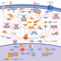
Signaling Pathway
Choose Selective ERK Inhibitors
Cell Data
| Cell Lines | Assay Type | Concentration | Incubation Time | Formulation | Activity Description | PMID |
|---|---|---|---|---|---|---|
| A375 | Function assay | Inhibition of ERK2 in human A375 cells harboring BRAF V600E mutant assessed as decrease in phosphorylated RSK levels, IC50 = 0.031 μM. | 28376306 | |||
| A375 | Function assay | 2 hrs | Inhibition of ERK1/2 in human A375 cells harboring B-RAF V600E mutant assessed as decrease in phospho-RSK level after 2 hrs by Cellomics ArrayScanTM VTI imaging analysis, IC50 = 0.14 μM. | 25977981 | ||
| A375 | Antiproliferative assay | 72 hrs | Antiproliferative activity against human A375 cells harboring B-RAF V600E mutant after 72 hrs by Cellomics ArrayScanTM VTI imaging analysis, IC50 = 0.18 μM. | 25977981 | ||
| SKCO1 | Antiproliferative assay | 72 hrs | Antiproliferative activity against human SKCO1 cells harboring KRAS G12V mutant after 72 hrs by CellTiter-Glo assay, IC50 = 0.356 μM. | 28038940 | ||
| BA/F3 | Antiproliferative assay | 72 hrs | Antiproliferative activity against mouse BA/F3 cells harboring KRAS G12D mutant after 72 hrs by CellTiter-Glo assay, IC50 = 0.468 μM. | 28038940 | ||
| SW620 | Antiproliferative assay | 72 hrs | Antiproliferative activity against human SW620 cells harboring KRAS G12V mutant after 72 hrs by CellTiter-Glo assay, IC50 = 0.499 μM. | 28038940 | ||
| AsPC1 | Antiproliferative assay | 72 hrs | Antiproliferative activity against human AsPC1 cells harboring KRAS G12D mutant after 72 hrs by CellTiter-Glo assay, IC50 = 0.849 μM. | 28038940 | ||
| NCI-H23 | Antiproliferative assay | 72 hrs | Antiproliferative activity against human NCI-H23 cells harboring KRAS G12C mutant after 72 hrs by CellTiter-Glo assay, IC50 = 1 μM. | 28038940 | ||
| SW156 | Antiproliferative assay | 72 hrs | Antiproliferative activity against human SW156 cells harboring wild type KRAS after 72 hrs by CellTiter-Glo assay, IC50 = 1.24 μM. | 28038940 | ||
| BA/F3 | Antiproliferative assay | 72 hrs | Antiproliferative activity against mouse BA/F3 cells after 72 hrs by CellTiter-Glo assay, IC50 = 2.231 μM. | 28038940 | ||
| A375 | Function assay | 2 hrs | Inhibition of ERK2 in human A375 cells harboring B-RAF V600E mutant assessed as decrease in phospho-ERK2 level after 2 hrs by Cellomics ArrayScanTM VTI imaging analysis, IC50 = 4.1 μM. | 25977981 | ||
| A375 | Function assay | Inhibition of ERK2 in human A375 cells harboring BRAF V600E mutant assessed as decrease in phosphorylated ERK2 levels, IC50 = 4.1 μM. | 28376306 | |||
| BA/F3 | Antiproliferative assay | 72 hrs | Antiproliferative activity against mouse BA/F3 cells harboring KRAS G12D mutant after 72 hrs in presence of IL-3 by CellTiter-Glo assay, IC50 = 8.608 μM. | 28038940 | ||
| Click to View More Cell Line Experimental Data | ||||||
Biological Activity
| Description | Ulixertinib (BVD-523, VRT752271) is a potent and reversible ERK1/ERK2 inhibitor with IC50 of <0.3 nM for ERK2. Phase 1. | ||||
|---|---|---|---|---|---|
| Targets |
|
| In vitro | ||||
| In vitro | In an A375 melanoma cell line containing a b-RAFV600E mutation, Ulixertinib reduces the levels of phosphorylated ERK2 (pERK) and of the phosphorylation of the downstream kinase RSK (pRSK) with IC50 of 4.1/0.14 μM, respectively. Ulixertinib also inhibits A375 cell proliferation with IC50 of 180 nM. [1] | |||
|---|---|---|---|---|
| Kinase Assay | ERK2 Rapidfire Mass Spectrometry Inhibition of Catalysis Assay | |||
| MEK U911-activated ERK2 protein is expressed and purified in-house. Enzyme and substrate solutions are made up in assay buffer consisting of 50 mM Tris (pH 7.5), 10 mM MgCl2, 0.1 mM EGTA, 10 mM DTT and 0.01% (v/v) CHAPS. 1.2 nM ERK2 protein is prepared in assay buffer and 10 µL is dispensed into each well of a polypropylene, 384-well plate containing test and reference control compounds. The compound plates had previously been dosed with a 12 point range from 100 µM down to 0.1 nM in order to calculate compound IC50s, with a total DMSO concentration in the assay of 1%. Following a 20 minute pre-incubation of enzyme and compound at room temperature, 10 µL of substrate solution is added consisting of 16 µM Erktide (IPTTPITTTYFFFK) and 120 µM ATP (measured Km) in assay buffer. The reaction is allowed to progress for 20 minutes at room temperature before being quenched by the addition of 80 µl 1% (v/v) formic acid. The assay plates are then run on the RapidFire Mass Spectrometry platform to measure substrate (unphosphorylated Erktide) and product (phosphorylated Erktide) levels. | ||||
| Cell Research | Cell lines | A375 cells | ||
| Concentrations | ~30 μM | |||
| Incubation Time | 72 h | |||
| Method | A375 cells are cultured in cell media composed of DMEM, 10% (v/v) Foetal Calf Serum and 1% (v/v) L-Glutamine. After harvesting, cells are dispensed into black, 384-well Costar plates to give 200 cells per well in a total volume of 40 µL cell media, and are incubated overnight at 37°C, 90% relative humidity and 5% CO2 in a rotating incubator. Test compounds and reference controls are dosed directly into the cell plates, into the inner 308 wells, using a Labcyte Echo 555 acoustic dispenser. The cells are dosed over a 12 point range from 30 µM down to 0.03 nM in order to calculate compound IC50s, with a total DMSO concentration in the assay of 0.3%. The cell plates are then incubated for 72 hours at 37°C. Cells were fixed and stained by the addition of 20 µL 12% formaldehyde in PBS/A (4% final concentration) and 1:2000 dilution of Hoechst 33342, with a 30 minute room temperature incubation, and then washed with PBS/A. A cell count is performed on the stained cell plates using a Cellomics ArrayScanTM VTI imaging platform. A Day 0 cell plate is also fixed, stained and read to generate a cell count baseline for determining compound cytotoxic effects as well as anti-proliferative effects. | |||
| Experimental Result Images | Methods | Biomarkers | Images | PMID |
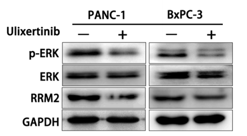
| Western blot | p-ERK / ERK / RRM2 |

|
28797284 | |
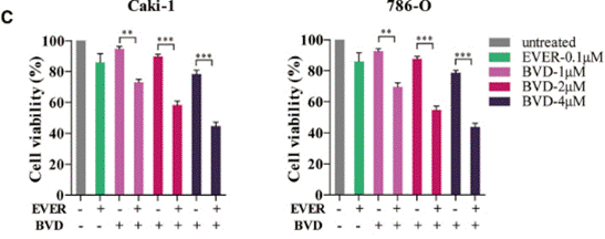
| Growth inhibition assay | Cell viability |

|
30771617 | |
| NCT Number | Recruitment | Conditions | Sponsor/Collaborators | Start Date | Phases |
|---|---|---|---|---|---|
| NCT04488003 | Active not recruiting | Advanced Solid Tumor|BRAF Gene Mutation|BRAF Gene Alteration|MEK Mutation|MEK Alteration|MAP2K1 Gene Mutation|MAP2K1 Gene Alteration|MAP2K2 Gene Mutation|MAP2K2 Gene Alteration |
BioMed Valley Discoveries Inc |
November 3 2020 | Phase 2 |
| NCT04145297 | Active not recruiting | Gastrointestinal Neoplasms |
University of Utah|BioMed Valley Discoveries Inc |
March 17 2020 | Phase 1 |
| NCT03698994 | Active not recruiting | Advanced Malignant Solid Neoplasm|Recurrent Ependymal Tumor|Recurrent Ewing Sarcoma|Recurrent Glioma|Recurrent Hepatoblastoma|Recurrent Histiocytic and Dendritic Cell Neoplasm|Recurrent Langerhans Cell Histiocytosis|Recurrent Malignant Germ Cell Tumor|Recurrent Malignant Solid Neoplasm|Recurrent Medulloblastoma|Recurrent Neuroblastoma|Recurrent Non-Hodgkin Lymphoma|Recurrent Osteosarcoma|Recurrent Peripheral Primitive Neuroectodermal Tumor|Recurrent Primary Malignant Central Nervous System Neoplasm|Recurrent Rhabdoid Tumor|Recurrent Rhabdomyosarcoma|Recurrent Soft Tissue Sarcoma|Refractory Ependymoma|Refractory Ewing Sarcoma|Refractory Glioma|Refractory Hepatoblastoma|Refractory Histiocytic and Dendritic Cell Neoplasm|Refractory Langerhans Cell Histiocytosis|Refractory Malignant Germ Cell Tumor|Refractory Malignant Solid Neoplasm|Refractory Medulloblastoma|Refractory Neuroblastoma|Refractory Non-Hodgkin Lymphoma|Refractory Osteosarcoma|Refractory Peripheral Primitive Neuroectodermal Tumor|Refractory Primary Malignant Central Nervous System Neoplasm|Refractory Rhabdoid Tumor|Refractory Rhabdomyosarcoma|Refractory Soft Tissue Sarcoma|Wilms Tumor |
National Cancer Institute (NCI) |
November 14 2018 | Phase 2 |
| NCT03417739 | Active not recruiting | Uveal Melanoma |
Dana-Farber Cancer Institute|BioMed Valley Discoveries Inc |
March 26 2018 | Phase 2 |
| NCT02994732 | Completed | Healthy |
BioMed Valley Discoveries Inc |
January 2017 | Phase 1 |
Chemical Information & Solubility
| Molecular Weight | 433.33 | Formula | C21H22Cl2N4O2 |
| CAS No. | 869886-67-9 | SDF | Download Ulixertinib (BVD-523) SDF |
| Smiles | CC(C)NC1=NC=C(C(=C1)C2=CNC(=C2)C(=O)NC(CO)C3=CC(=CC=C3)Cl)Cl | ||
| Storage (From the date of receipt) | |||
|
In vitro |
DMSO : 86 mg/mL ( (198.46 mM); Moisture-absorbing DMSO reduces solubility. Please use fresh DMSO.) Ethanol : 86 mg/mL Water : Insoluble |
Molecular Weight Calculator |
|
In vivo Add solvents to the product individually and in order. |
In vivo Formulation Calculator |
||||
Preparing Stock Solutions
Molarity Calculator
In vivo Formulation Calculator (Clear solution)
Step 1: Enter information below (Recommended: An additional animal making an allowance for loss during the experiment)
mg/kg
g
μL
Step 2: Enter the in vivo formulation (This is only the calculator, not formulation. Please contact us first if there is no in vivo formulation at the solubility Section.)
% DMSO
%
% Tween 80
% ddH2O
%DMSO
%
Calculation results:
Working concentration: mg/ml;
Method for preparing DMSO master liquid: mg drug pre-dissolved in μL DMSO ( Master liquid concentration mg/mL, Please contact us first if the concentration exceeds the DMSO solubility of the batch of drug. )
Method for preparing in vivo formulation: Take μL DMSO master liquid, next addμL PEG300, mix and clarify, next addμL Tween 80, mix and clarify, next add μL ddH2O, mix and clarify.
Method for preparing in vivo formulation: Take μL DMSO master liquid, next add μL Corn oil, mix and clarify.
Note: 1. Please make sure the liquid is clear before adding the next solvent.
2. Be sure to add the solvent(s) in order. You must ensure that the solution obtained, in the previous addition, is a clear solution before proceeding to add the next solvent. Physical methods such
as vortex, ultrasound or hot water bath can be used to aid dissolving.
Tech Support
Answers to questions you may have can be found in the inhibitor handling instructions. Topics include how to prepare stock solutions, how to store inhibitors, and issues that need special attention for cell-based assays and animal experiments.
Tel: +1-832-582-8158 Ext:3
If you have any other enquiries, please leave a message.
* Indicates a Required Field
Tags: buy Ulixertinib (BVD-523) | Ulixertinib (BVD-523) supplier | purchase Ulixertinib (BVD-523) | Ulixertinib (BVD-523) cost | Ulixertinib (BVD-523) manufacturer | order Ulixertinib (BVD-523) | Ulixertinib (BVD-523) distributor


