
Why is Tamoxifen failing to inhibit MCF-7 cell growth in vitro compared to 4-hydroxytamoxifen?
Quick Answer
Recommended ApproachUse the active metabolite 4-hydroxytamoxifen rather than parent tamoxifen at 1 to 10 micromolar in phenol red-free medium supplemented with charcoal-stripped serum to ensure potent estrogen receptor engagement.
Common CausesParent tamoxifen failed to inhibit MCF-7 cell proliferation because cultured breast cancer cells lack the hepatic cytochrome P450 enzymes required to bioactivate tamoxifen into its high-affinity active metabolites.
How Should It Be Done Correctly?
In Vitro Endocrine Sensitivity Assay Specifications
| Parameter | Recommended Specification | Operational Note |
|---|---|---|
| Active Compound | 4-Hydroxytamoxifen (4-OHT / 4OHtam) | Active metabolite bypassing hepatic CYP requirement [1, 2, 3] |
| Stock Concentration | 10 mM in anhydrous DMSO | Aliquot and store at -80°C (1-year shelf life) or -20°C (1 month) |
| In Vitro Working Concentration | 1 to 10 µM | Induces concentration-dependent cell cycle arrest and apoptosis [3, 4] |
| Culture Medium | Phenol red-free MEM, DMEM, or RPMI 1640 | Eliminates intrinsic estrogenic background of phenol red [1, 2, 5] |
| Serum Supplement | 3% to 5% Charcoal-stripped FBS (csFBS) | Depletes endogenous steroid hormones before antiestrogen challenge [2, 5] |
| Final Solvent Concentration | ≤ 0.1% (v/v) DMSO | Prevents vehicle-induced non-specific membrane toxicity |
| Pre-treatment Depletion | 24 to 72 hours in csFBS medium | Clears basal receptor occupancy and synchronizes cells [2, 5] |
| Assay Duration | 48 to 72 hours post-treatment | Required window to observe full apoptotic and metabolic arrest [1, 3, 5] |
Operational Procedure for In Vitro Antiestrogen Testing
- Reagent Preparation and Solubilization: Dissolve 4-hydroxytamoxifen (or tamoxifen powder stored desiccated at -20°C) in high-grade anhydrous DMSO to establish a 10 mM stock solution. Vortex thoroughly until completely dissolved. Prepare single-use aliquots to prevent hygroscopic moisture absorption and repeated freeze-thaw degradation, then immediately freeze at -80°C. Working dilutions should be prepared fresh directly before treatment.
- Hormone Depletion Phase: Seed MCF-7 cells at 5,000 to 10,000 cells per well in 96-well format (or proportionally in 6-well/12-well formats) [5]. Allow cells to attach overnight. At 24 to 72 hours prior to drug administration, aspirate standard growth medium, wash monolayers gently with pre-warmed PBS, and introduce phenol red-free basal medium containing 3% to 5% charcoal-stripped FBS (csFBS) [2, 5]. This ensures complete clearance of exogenous estrogens present in regular serum.
- Drug Dosing and Dilution Workflow: Prepare serial dilutions of 4-OHT in pre-warmed, hormone-depleted medium. Dispense the working dilution to achieve final concentrations spanning 1 µM to 10 µM [3, 4]. Ensure the final vehicle concentration remains constant across all test groups and matching vehicle control wells (≤ 0.1% v/v DMSO). Gently agitate plates to ensure homogeneous dispersion.
- Incubation and Endpoint Evaluation: Maintain cells at 37°C in a humidified 5% CO2 incubator for 48 to 72 hours without disturbing the microenvironment [2, 3, 5]. Assess cell viability and proliferation using MTS, MTT, or BrdU incorporation assays [4, 5, 6]. Complement metabolic viability measurements with flow-cytometry-based apoptosis assays (e.g., Annexin V/PI) to distinguish cytostatic G1 arrest from apoptotic induction [3].
What Went Wrong?
Copy code to add this image to your page Copy Code
In Vitro Administration of Parent Tamoxifen Prodrug
The fundamental operational error was dosing in vitro cell cultures directly with unmodified parent tamoxifen expecting equivalent efficacy to clinical responses. Cell-based assays lack the anatomical and multi-tissue pharmacokinetic context of systemic administration. Applying parental tamoxifen to cell culture plates fails to elicit substantial cytostatic or cytotoxic effects unless raised to supraphysiological, non-specific toxic concentrations (≥ 10–15 µM), at which point cell death occurs independent of estrogen receptor regulation [1, 6].
Absence of Cytochrome P450 Bioactivation
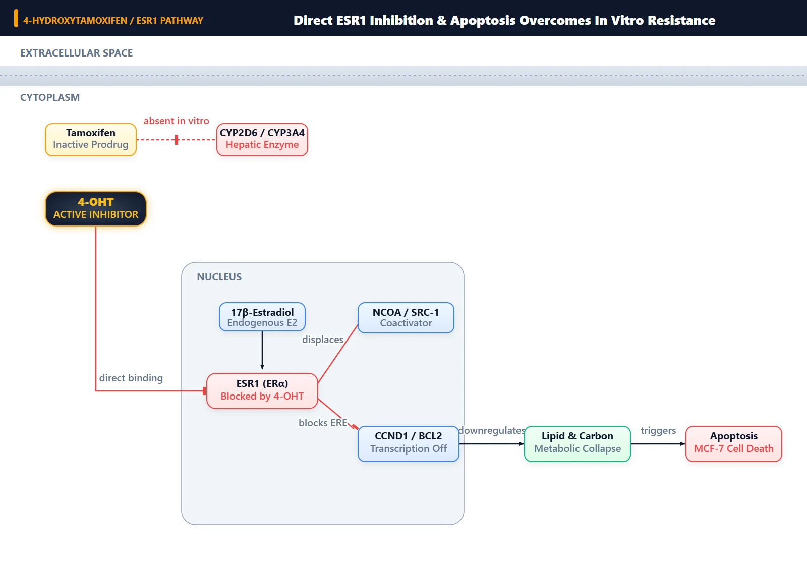
The proximate cause of the experimental failure is that MCF-7 breast adenocarcinoma cells do not express the requisite complement of hepatic cytochrome P450 (CYP) enzymes, most notably CYP2D6 and CYP3A4. In human patients, tamoxifen serves as a prodrug that undergoes extensive phase I hepatic biotransformation into active derivatives, including 4-hydroxytamoxifen (4-OHT) and N-desmethyl-4-hydroxytamoxifen (endoxifen) [1, 2]. Cultured MCF-7 monolayers cannot convert parent tamoxifen into these active metabolites, leaving the cell exposed almost entirely to the low-affinity parent compound.
Target Engagement Disparity and Transcriptional Failure
The underlying molecular mechanism centers on the differential binding affinity for estrogen receptor alpha (ERα, encoded by ESR1) and subsequent transcriptional repression. Parent tamoxifen possesses weak relative binding affinity for ERα, whereas 4-hydroxytamoxifen displays an affinity nearly 100- to 1000-fold higher, comparable to that of endogenous 17β-estradiol (E2) [1, 4]. When 4-OHT enters an ERα-positive MCF-7 cell, it competitively displaces estrogen from the ligand-binding domain (LBD), stabilizing an antagonistic receptor conformation that inhibits coregulator recruitment (such as SRC-3) and silences pro-survival target genes [1, 4].
Furthermore, high-affinity engagement by 4-OHT triggers widespread metabolic rewiring in MCF-7 cells, specifically downregulating ATP-citrate lyase (ACLY) and disrupting central carbon and de novo lipid metabolism, which collapses the mitochondrial membrane potential and activates caspase-dependent apoptosis [3]. Because parental tamoxifen cannot achieve adequate ERα occupancy at physiological concentrations, it fails to alter global gene expression, repress essential keratins such as KRT6, or induce mitochondrial apoptosis [1, 3].
How Can I Avoid This in Future Experiments?
To ensure consistent and reproducible results in endocrine response assays, always verify whether experimental compounds require metabolic bioactivation before testing them in monolayer culture. For any SERM or antiestrogenic study in MCF-7, T47D, or ZR-75-1 cell lines, select active metabolites (4-hydroxytamoxifen or endoxifen) as the primary pharmacological probes [1, 2, 3].
Additionally, standard basal formulations contain phenol red, which functions as a weak estrogen mimic capable of competing with low-affinity SERMs. Always pair active metabolite dosing with certified phenol red-free medium and charcoal-dextran-stripped fetal bovine serum to eliminate background estrogen receptor stimulation [2, 5]. Maintain vehicle DMSO below 0.1% to avoid solvent artifacts on membrane permeability and cell viability.
References
- [1]Helland T, et al. PloS one. 2015 Jan;10(4):e0122339.[IF: 3.057]
- [2]Kinga Linowiecka, Justyna Szpotan, Marlena Godlewska, Damian Gaweł, Ewelina Zarakowska, Daniel Gackowski, Anna A. Brożyna and Marek Foksiński, et al. International journal of molecular sciences..
- [3]Kazuki Nishimoto, et al. Scientific reports..
- [4]Devulapally R, et al. Molecular pharmaceutics. 2015 Jun;12(6):2080-92.[IF: 4.342]
- [5]Unknown, et al. International journal of molecular sciences..
- [6]Chang YC, et al. Frontiers in cell and developmental biology. 2020 Jan;8:485.[IF: 6.684]