
Why did Cre-ERT2 recombination fail in brain tissue despite administering 2 mg Tamoxifen daily?
Quick Answer
Recommended ApproachAdminister tamoxifen at 75–100 mg/kg daily via intraperitoneal injection for 3 to 5 consecutive days and allow at least 7 to 21 days post-injection before tissue collection.
Common CausesRecombination failed because a single daily dose or insufficient consecutive administration days cannot sustain the threshold cerebral concentrations of the active metabolite 4-hydroxytamoxifen required to induce Cre nuclear translocation across floxed alleles in the brain.
How Should It Be Done Correctly?
Recommended Dosing and Reagent Parameters
Key chemical properties, formulation parameters, and in vivo dosing regimens for inducing robust Cre-ERT2 recombination in murine neural tissue. [1, 2]
| Parameter | Specification / Recommended Protocol | Target Tissue / Biological Context |
|---|---|---|
| Stock Solid Storage | -20°C, dry powder, desiccated (shelf life: 3 years) | Chemical reagent integrity |
| Stock Solvent Storage | DMSO stock at -80°C (1 year) or -20°C (1 month) | Aliquoted working stocks |
| DMSO Solubility | Up to 74 mg/mL (recommended stock: 10 mM) | Reconstitution verification |
| Aqueous Solubility | <1 mg/mL (insoluble in water/saline) | Vehicle selection constraint |
| In Vivo Dosing Vehicle | Corn oil or sunflower oil with 5–10% absolute ethanol | Intraperitoneal injection solution |
| In Vivo Working Dosage | 75–100 mg/kg body weight (approx. 1.5–2.0 mg/mouse) | Adult C57BL/6 background mice |
| Injection Frequency | Once daily for 3 consecutive days | Cerebellum and highly accessible loci |
| Injection Frequency | Once daily for 5 consecutive days | Cerebral cortex and tightly chromatin-packed loci |
| Post-Injection Washout | 7 to 21 days post-final injection | Reporter maturation and drug clearance |
Standardized Preparation and Administration Protocol
- Vehicle Preparation and Dissolution: Formulate the lipophilic injection medium by dissolving powdered tamoxifen in sterile corn oil or sunflower oil pre-supplemented with absolute ethanol [2, 3]. Heat the mixture in an orbital shaker or water bath at 55–65°C for 1–2 hours, vortexing at 15-minute intervals until no microcrystalline particulates remain.
- Quality Inspection: Inspect the solubilized solution against a dark background under bright light to verify complete optical clarity. Filter the solution through a 0.22 µm PTFE syringe filter to maintain sterility without drug sorption.
- Consecutive Injection Schedule: Administer the vehicle solution intraperitoneally at calibrated volume loads (typically 100 µL for a 20 g mouse) using a 27-gauge needle [2]. Repeat the injection at the same circadian hour each day across consecutive days rather than relying on a single or fragmented application [2].
- Post-Administration Maturation Interval: House the treated animals under standard maintenance conditions for a recovery window after the final injection before tissue harvest [2, 4]. This interval permits nuclear translocation of Cre recombinase, genomic strand cleavage, loxP site excision, and adequate accumulation of the downstream reporter protein or decay of targeted floxed transcripts [2].
What Went Wrong?
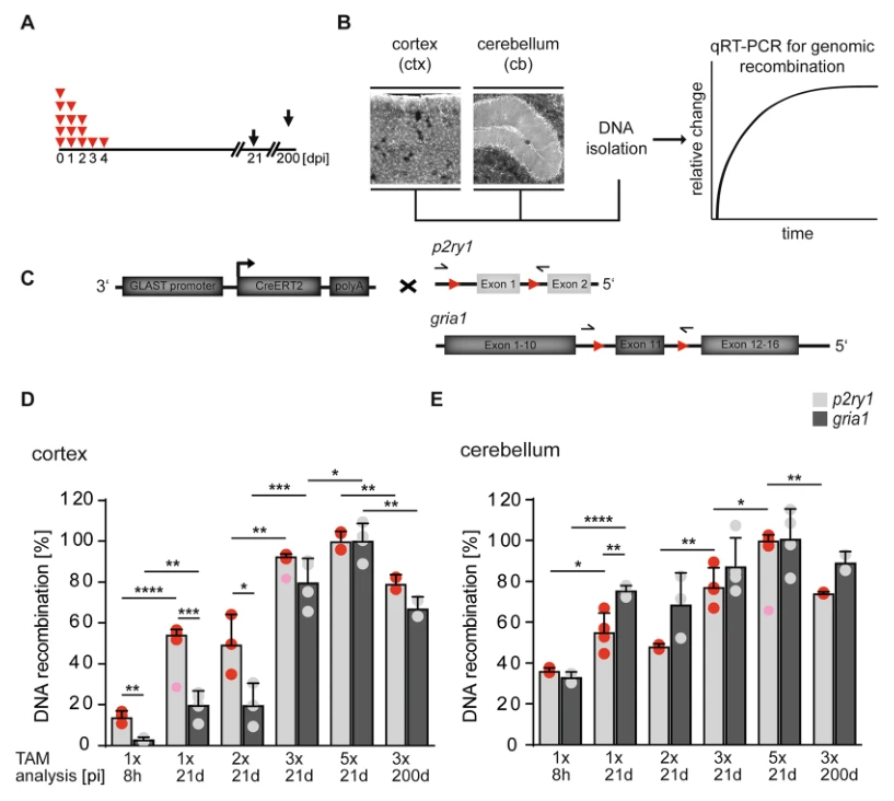
Copy code to add this image to your page Copy Code
Insufficient Administration Duration and Cumulative Exposure
The most frequent operational oversight in conditional knockout studies in the central nervous system is administering tamoxifen as an isolated 1-day dose or over an inadequate sequence of consecutive days [2]. Although 2 mg matches the standard daily mass dose for an adult mouse (representing 80–100 mg/kg), achieving robust recombination in the brain requires sustaining target engagement over time rather than providing a single acute bolus [2]. Single injections typically induce nominal or undetectable recombination at tightly regulated neural loci, whereas multi-day consecutive regimens achieve near-maximal genomic excision [2].
Rapid Metabolic Clearance Across the Blood-Brain Barrier
Following systemic intraperitoneal delivery, parent tamoxifen and its hydroxylated derivatives readily cross the blood-brain barrier, reaching peak cerebral tissue concentrations around 8 hours post-injection [2]. However, cerebral elimination is rapid: parent drug concentrations decline sharply within 24 hours, and effective active metabolites are largely depleted within several days [2]. A single administration fails to maintain the critical intra-parenchymal concentration threshold needed to achieve broad coverage across neural cell populations [2]. Multi-day consecutive dosing is required to establish prolonged tissue enrichment and delay drug clearance, ensuring sustained enzymatic engagement in cerebral structures [2].
Inadequate Cre-ERT2 Nuclear Retention and Locus-Specific Accessibility Constraints
Cre-ERT2 remains tethered in the cytoplasm by heat shock proteins in the absence of ligand. Induction requires the active metabolite 4-hydroxytamoxifen (4-OH-TAM) or endoxifen to bind the mutated estrogen receptor ligand-binding domain (ERT2), triggering conformational dissociation and nuclear translocation [2, 4]. Because tamoxifen conversion yields 4-OH-TAM in relatively low proportions compared to parent drug levels in the brain (approximately 4% to 6% of parent drug at peak hours) [2], an isolated dose results in transient, short-lived nuclear occupancy. Furthermore, different genomic loci display distinct steric accessibility; heterochromatin-embedded floxed loci (such as Gria1 or P2ry1) demand significantly higher and more persistent nuclear Cre concentrations than permissive reporter loci (such as Rosa26) to achieve productive recombination [2].
How Can I Avoid This in Future Experiments?
To ensure consistent recombination in future central nervous system experiments, establish a validated multi-day intraperitoneal regimen tailored to the specific anatomical brain region and target locus [2]. For cortical astrocytic and neuronal targets, implement a full 5-consecutive-day protocol, while 3 consecutive days is typically sufficient for cerebellar populations [2]. Ensure that the floxed allele recombines efficiently by validating recombination at the genomic DNA level via quantitative PCR using primers spanning the flanking loxP sites, rather than relying exclusively on fluorescent reporter signals [2].
Always prepare tamoxifen fresh or aliquot single-use working volumes protected from light, because repeated warming or freeze-thaw cycles precipitate the compound and degrade dosing consistency. Finally, incorporate an adequate post-injection maturation window of at least 14 to 21 days before tissue collection to allow complete phenotypic protein turnover and clearance of any transient cellular stress induced by the drug [2, 3].
References
- [1]Jovanovic J, et al. bioRxiv : the preprint server for biology. 2025 Jan.
- [2]Jahn HM, et al. Scientific reports. 2018 Apr;8(1):5913.[IF: 4.011]
- [3]Denk F, et al. Acta neuropathologica communications. 2015 Nov;3:74.[IF: 5.414]
- [4]Falke LL, et al. Journal of cell communication and signaling. 2017 Jun;11(2):205-211.[IF: 2.516]