
Why does Tamoxifen show severe loss of free concentration in untreated polystyrene 96-well plates?
Quick Answer
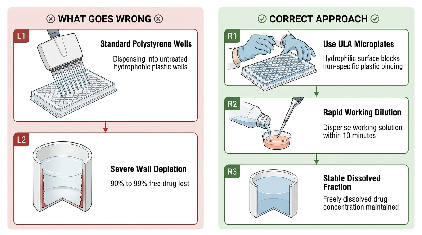
Recommended ApproachTo maintain intended free concentrations of Tamoxifen, perform in vitro assays in ultra-low attachment (ULA) or hydrophilic-passivated plates, minimize working solution hold times, and account for extensive serum protein binding and surface sorption in dosimetry calculations.
Common CausesSevere loss of free Tamoxifen occurs because its extreme lipophilicity drives rapid, non-specific adsorption onto hydrophobic untreated polystyrene surfaces, which combines with high serum protein binding to strip freely dissolved drug from the aqueous phase.
How Should It Be Done Correctly?
Copy code to add this image to your page Copy Code
Vessel Selection and Handling Specifications
Operational parameters and vessel compatibility guidelines for preventing non-specific loss of Tamoxifen in microplate-based assays.
Step-by-Step Dosing and Incubation Protocol
-
Reconstitute and Aliquot Stock Solution Dissolve solid Tamoxifen powder in high-grade anhydrous DMSO to establish a 10 mM stock solution. Vigorously vortex the vial for 60 seconds until the solution is completely clear and free of particulate matter. Immediately divide the stock into single-use low-retention microcentrifuge tubes to prevent repeated freeze-thaw cycles and ambient moisture absorption. Store these working aliquots at -20°C for up to 1 month or -80°C for up to 1 year, thawing only the required volume immediately prior to experiment setup.
-
Passivate Plate Surfaces or Deploy Low-Binding Consumables Do not expose aqueous Tamoxifen solutions to standard untreated or non-tissue-culture-treated polystyrene. Use pre-sterilized microplates engineered with a covalently bonded hydrogel layer (such as standard commercially available ultra-low attachment surfaces). If low-binding plates are unavailable, pre-incubate untreated polystyrene wells with complete culture medium containing serum proteins or a sterile 0.1% bovine serum albumin (BSA) solution in PBS for at least 1 hour at 37°C to block active non-specific binding sites before introducing the test compound.
-
Prepare Fresh Intermediate Dilutions in Warm Matrix Warm the culture medium or assay buffer to 37°C prior to compounding. Dilute the 10 mM DMSO stock into the medium using rapid, stepwise mixing to ensure immediate dispersal without localized phase separation or precipitation. Keep final vehicle concentrations strictly at or below 0.1%–0.5% (v/v) DMSO across all test wells. Never prepare intermediate aqueous dilutions in plain polystyrene reservoirs hours in advance; compound solutions must be dispensed into the assay vessel within 5–10 minutes of preparation to curb wall depletion.
-
Apply Realistic Dosimetry Calculations When correlating nominal concentrations to biological responses, calibrate for true free concentrations (Cfᵣₑₑ) by running parallel cell-free recovery measurements using HPLC-UV or LC-MS/MS [1, 2]. Integrate mass balance considerations that partition the total substance into plastic-adsorbed, protein-bound, intracellular, and freely dissolved fractions to prevent severe underestimation of drug potency [1].
| Parameter / Condition | Specification / Recommended Setting | Purpose / Operational Context |
|---|---|---|
| Recommended Stock Concentration | 10 mM in anhydrous DMSO | Stable concentrated stock minimizing vehicle volume in assays |
| Stock Solvent Solubility | 74 mg/mL in DMSO; 74 mg/mL in Ethanol | Complete dissolution of stock powder before dilution |
| Aqueous Solubility Limit | < 1 mg/mL in water (practically insoluble) | Prevents micelle formation or micro-precipitation in buffer |
| Powder Storage | -20°C, sealed desiccated (shelf life: 3 years) | Preserves chemical integrity of bulk compound |
| Solution Storage | -80°C (1 year) or -20°C (1 month) | Prevents vehicle evaporation and freeze-thaw solute fallout |
| Primary Assay Plate Plastic | Ultra-low attachment (ULA) or PEG-grafted cyclic olefin | Eliminates hydrophobic plastic binding sites |
| Alternative Plate Pre-treatment | Pre-coating with 0.1% BSA or 0.05% Pluronic F-127 | Saturates residual hydrophobic adsorption loci |
| Serum Fraction Dosimetry Adjustment | Account for ~99% protein binding (fᵤ ≈ 1%) | Compensates for medium protein sequestration in dose-response [1] |
What Went Wrong?
Inappropriate Plate Chemistry and Hydrophobic Adsorption
Standard untreated polystyrene consists of unmodified, non-polar polymer chains lacking the oxygen-containing hydrophilic functional groups introduced during tissue-culture (TC) gas-plasma treatment or specialized hydrogel passivation coatings. Dispensing aqueous solutions of Tamoxifen directly into untreated polystyrene 96-well plates exposes the compound to a vast surface area of exposed phenyl groups. Due to the absence of a hydrophilic barrier or blocking agents, dissolved Tamoxifen molecules partition out of the aqueous phase and adhere tenaciously to the well walls and bottoms, resulting in immediate and massive depletion from the liquid column.
Compound Depletion from the Freely Dissolved Phase
Tamoxifen is an exceptionally lipophilic small molecule (calculated log KOW > 6) exhibiting negligible aqueous solubility (< 1 mg/mL) [1, 2]. When introduced into an aqueous microplate environment, the thermodynamic drive to escape unfavorable polar interactions with water pushes Tamoxifen molecules toward any available hydrophobic sink. In untreated polystyrene vessels, this sink is primarily the bare plastic surface, followed in culture systems by rapid binding to medium proteins [1]. The unbound fraction (fᵤ) of Tamoxifen in standard serum-containing media can drop to approximately 1% or lower [1]. Consequently, free drug concentrations available to interact with molecular targets in vitro can plummet by 90%–99% compared to the nominal concentration (CNₒₘ) added by the investigator [1].
Molecular Partitioning and Sequestration Dynamics
The primary driving force behind plastic loss is non-specific hydrophobic interaction combined with van der Waals and pi--pi stacking between the aromatic rings of Tamoxifen and the polystyrene backbone. Unlike polar chemicals that remain stable in the bulk aqueous phase, Tamoxifen establishes a mass-balance equilibrium heavily skewed toward the solid-liquid interface [1]. In cellular setups, this surface loss is compounded by rapid trans-membrane diffusion and lysosomal trapping—driven by Tamoxifen's weakly basic tertiary amine group, which becomes protonated in acidic organelles—leading to severe cellular and matrix sequestration that leaves negligible unbound compound in the active supernatant [1].
How Can I Avoid This in Future Experiments?
To prevent catastrophic free concentration loss in future screening runs, replace all sample contact plastics with verified low-retention materials. This includes using low-binding polypropylene pipette tips, glass-coated or fluoropolymer microplate inserts, and hydrogel-coated ULA plates across the entire liquid handling chain.
Always incorporate serum-free or carrier-supplemented controls when characterizing novel target affinities or cytotoxicity curves. For highly lipophilic drugs like Tamoxifen, relying strictly on nominal dosing concentrations creates extreme systematic error; quantifying actual free concentrations in conditioned media via equilibrium dialysis or solid-phase microextraction ensures reproducible, physiologically meaningful concentration-response profiles [1].