-
 Australia
-
 Austria
-
 Belgium
-
 Brazil
-
 Canada
-
 China
China
-
 Czech Republic
-
 Denmark
-
 Finland
-
 France
-
 Germany
-
 Greece
-
 Hong Kong
-
 Hungary
-
 Iceland
-
 India
India
-
 Ireland
-
 Israel
-
 Italy
Italy
-
 Japan
Japan
-
 Korea
Korea
-
 Luxembourg
-
 Malaysia
-
 Netherlands
-
 New Zealand
-
 Norway
-
 Poland
-
 Qatar
Qatar
-
 Romania
-
 Saudi Arabia
-
 Singapore
-
 Spain
Spain
-
 Sweden
-
 Switzerland
-
 Taiwan
-
 Turkey
-
 United Kingdom
-
 United States
research use only
PHA-767491 HCl CDK inhibitor
Cat.No.S2742
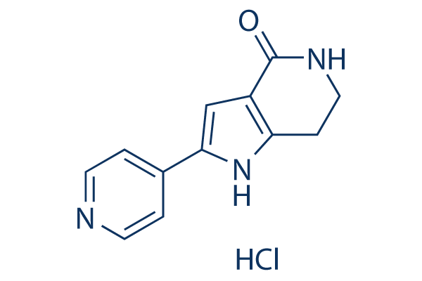
Chemical Structure
Molecular Weight: 249.7
Quality Control
Cell Culture, Treatment & Working Concentration
| Cell Lines | Assay Type | Concentration | Incubation Time | Formulation | Activity Description | PMID |
|---|---|---|---|---|---|---|
| human SF268 cells | Proliferation assay | 72 h | Antiproliferative activity against p53 deficient human SF268 cells after 72 hrs, IC50=0.86 μM | |||
| human HCT116 cells | Proliferation assay | 72 h | Antiproliferative activity against human HCT116 cells expressing p53 gene after 72 hrs by proliferative assay, IC50=0.97 μM | |||
| human HCT16 cells | Proliferation assay | 72 h | Antiproliferative activity against human HCT16 cells after 72 hrs by luciferase based assay, IC50=1 μM | |||
| human SW403 cells | Proliferation assay | 72 h | Antiproliferative activity against human SW403 cells after 72 hrs by luciferase based assay, IC50=1 μM | |||
| human A2780 cells | Proliferation assay | 72 h | Antiproliferative activity against human A2780 cells expressing p53 gene after 72 hrs by proliferative assay, IC50=1.07 μM | |||
| human SW48 cells | Proliferation assay | 72 h | Antiproliferative activity against human SW48 cells after 72 hrs by luciferase based assay, IC50=1.2 μM | |||
| human MCF7 cells | Proliferation assay | 72 h | Antiproliferative activity against human MCF7 cells after 72 hrs by luciferase based assay, IC50=1.3 μM | |||
| human U2OS cells | Proliferation assay | 72 h | Antiproliferative activity against human U2OS cells expressing p53 gene after 72 hrs by proliferative assay, IC50=1.49 μM | |||
| human COLO205 cells | Proliferation assay | 72 h | Antiproliferative activity against human COLO205 cells after 72 hrs by luciferase based assay, IC50=1.5 μM | |||
| human OVCAR8 cells | Proliferation assay | 72 h | Antiproliferative activity against p53 deficient human OVCAR8 cells after 72 hrs by proliferative assay, IC50=1.56 μM | |||
| human L363 cells | Proliferation assay | 72 h | Antiproliferative activity against human L363 cells after 72 hrs by luciferase based assay, IC50=1.6 μM | |||
| human NHDF cells | Proliferation assay | 72 h | Antiproliferative activity against human NHDF cells after 72 hrs by luciferase based assay, IC50=1.6 μM | |||
| human NCI-H929 cells | Proliferation assay | 72 h | Antiproliferative activity against human NCI-H929 cells after 72 hrs by luciferase based assay, IC50=1.8 μM | |||
| human SF539 cells | Proliferation assay | 72 h | Antiproliferative activity against human SF539 cells expressing p53 gene after 72 hrs by proliferative assay, IC50=2.34 μM | |||
| human SW480 cells | Proliferation assay | 72 h | Antiproliferative activity against p53 deficient human SW480 cells after 72 hrs by proliferative assay, IC50=2.67 μM | |||
| human NCI60 cells | Proliferation assay | Antiproliferative activity against human NCI60 cells, IC50=3.1 μM | ||||
| human Jurkat cells | Proliferation assay | 72 h | Antiproliferative activity against p53 deficient human Jurkat cells after 72 hrs by proliferative assay, IC50=3.2 μM | |||
| human HCT15 cells | Proliferation assay | 72 h | Antiproliferative activity against human HCT15 cells expressing p53 gene after 72 hrs by proliferative assay, IC50=3.81 μM | |||
| human OPM2 cells | Proliferation assay | 72 h | Antiproliferative activity against human OPM2 cells after 72 hrs by luciferase based assay, IC50=4.5 μM | |||
| human HT-29 cells | Proliferation assay | 72 h | Antiproliferative activity against human HT-29 cells after 72 hrs by luciferase based assay, IC50=5 μM | |||
| human K562 cells | Proliferation assay | 72 h | Antiproliferative activity against p53 deficient human K562 cells after 72 hrs, IC50=5.87 μM | |||
| U937 cells | Function assay | Inhibition of TNFalpha production in U937 cells, IC50=19 μM | ||||
| human HeLa cells | Function assay | 5 μM | 24 h | Induction of apoptosis in human HeLa cells assessed as appearance of PARP at 5 uM after 24 hrs | ||
| NHDF | Function assay | 5 μM | 16 h | Induction of cell cycle arrest in thymidine deficient NHDF assessed as DNA synthesis in S-phase at 5 uM after 16hrs FACS analysis in presence of serum | ||
| Click to View More Cell Line Experimental Data | ||||||
Solubility
|
In vitro |
DMSO
: 24 mg/mL
(96.11 mM)
Water : Insoluble Ethanol : Insoluble |
Molarity Calculator
|
In vivo |
|||||
In vivo Formulation Calculator (Clear solution)
Step 1: Enter information below (Recommended: An additional animal making an allowance for loss during the experiment)
Step 2: Enter the in vivo formulation (This is only the calculator, not formulation. Please contact us first if there is no in vivo formulation at the solubility Section.)
Calculation results:
Working concentration: mg/ml;
Method for preparing DMSO master liquid: mg drug pre-dissolved in μL DMSO ( Master liquid concentration mg/mL, Please contact us first if the concentration exceeds the DMSO solubility of the batch of drug. )
Method for preparing in vivo formulation: Take μL DMSO master liquid, next addμL PEG300, mix and clarify, next addμL Tween 80, mix and clarify, next add μL ddH2O, mix and clarify.
Method for preparing in vivo formulation: Take μL DMSO master liquid, next add μL Corn oil, mix and clarify.
Note: 1. Please make sure the liquid is clear before adding the next solvent.
2. Be sure to add the solvent(s) in order. You must ensure that the solution obtained, in the previous addition, is a clear solution before proceeding to add the next solvent. Physical methods such
as vortex, ultrasound or hot water bath can be used to aid dissolving.
Chemical Information, Storage & Stability
| Molecular Weight | 249.7 | Formula | C12H11N3O.HCl |
Storage (From the date of receipt) | |
|---|---|---|---|---|---|
| CAS No. | 942425-68-5 | Download SDF | Storage of Stock Solutions |
|
|
| Synonyms | CAY10572, NMS 1116354 | Smiles | C1CNC(=O)C2=C1NC(=C2)C3=CC=NC=C3.Cl | ||
Mechanism of Action
| Features |
The first inhibitor that directly affects the mechanisms controlling initiation as opposed to elongation in DNA replication.
|
|---|---|
| Targets/IC50/Ki |
Cdc7
(Cell-free assay) 10 nM
CDK9
(Cell-free assay) 34 nM
GSK-3β
(Cell-free assay) 220 nM
CDK2
(Cell-free assay) 240 nM
CDK1
(Cell-free assay) 250 nM
CDK5
(Cell-free assay) 460 nM
MK2
(Cell-free assay) 470 nM
PLK1
(Cell-free assay) 980 nM
|
| In vitro |
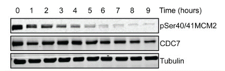
PHA-767491 displays approximately 20-fold selectivity for Cdk1, Cdk2 and GSK3-β, 50-fold selectivity for MK2 and Cdk5 and 100-fold selectivity for PLK1 and CHK2. PHA-767491 inhibits cell proliferation in a variety of human cell lines with IC50 of 0.86 μM for SF-268 to 5.87 μM for K562, and significantly induces apoptosis in a p53-independent manner in almost all cell lines in contrast with 5-FU which only works in a few of cell lines. Unlike current DNA synthesis inhibitors, PHA-767491 treatment at 5 μM blocks the initiation of DNA replication but not replication fork progression, due to specific inhibition of Cdc7 kinase and Mcm2 phosphorylation at the Cdc7-dependent Ser40 site. The up-regulated Mcl-1 levels in ABT-737-resistant OCI-LY1 and SU-DHL-4 cells can be significantly decreased by PHA-767491 treatment at 3 μM possibly due to the inhibition of Cdk9, leading to the restoration of the sensitivity to ABT-737. The direct mitochondrial dependent pro-apoptosis effect of PHA-767491 is also observed when applied at 1 μM in quiescent chronic lymphocytic leukemia (CLL) cells through the similar mechanism with EC50 of 0.34-0.97 μM. While in proliferating CLL cells stimulated by CD154 and interleukin-4, PHA-767491 treatment at 5 μM abolishes DNA synthesis by inhibiting Cdc7 rather than triggering cell death. |
| Kinase Assay |
In vitro kinase assays
|
|
The inhibition of Cdc7 and Cdk9 by PHA-767491 (IC50) is determined using the strong anion exchanger (Dowex 1-X8 resin, formate form)-based assay. For each enzyme, the absolute Km values for ATP and the specific substrate are initially determined, and each assay is then run at optimized ATP/33P-γ-ATP mix (2Km) and substrate (5Km) concentrations. Cdc7 kinase assay is performed in a buffer containing 50 mM Hepes pH 7.9, 15 mM MgCl2, 2 mM β- glycerylphosphate, 0.2 mg/mL BSA, 1 mM DTT, 3 μM Na3VO4, 2Km ATP/33P-γ-ATP mix, 5Km Mcm2 (aa 10-294), 37 nM of recombinant Cdc7/Dbf4 and increasing concentration of PHA-767491 in a final volume of 30 μL, and incubated for 1 hour at 25 °C. Cdk9 kinase assay is performed using 50 nM of recombinant Cdk9/cyclin T in 50 mM HEPES pH 7.5, 10 mM MgCl2, 1 mM DTT, 3 μM Na3VO4, 2Km ATP/33P-γ-ATP mix, 5Km RNA polymerase CDT peptide and increasing concentration of PHA-767491 in a final volume of 30 μL, and incubated for 1 hour at 25 °C. After incubation, an amount of 150 μL of resin/formate (pH 3.0) is added to stop the reaction and capture unreacted 33P-γ-ATP, separating it from the phosphorylated substrate in solution. After 1 hour of rest, a volume of 50 μL supernatant is transferred to Optiplate 96-well plates. After the additon of 150 μL of Microscint 40, the radioactivity is counted in the TopCount.
|
|
| In vivo |
Administration of PHA-767491 twice a day for 5 days significantly inhibits the growth of HL60 xenograft in a dose-dependent manner with TGI of 50% and 92% at dose of 20 mg/kg and 30 mg/kg, respectively, the effect of which is also marked in A2780, Mx-1, and HCT-116 xenograft models as well as the mammary carcinomas, and correlates with Cdc7 inhibition and subsequently decreased phosphorylation of Mcm2 at the Cdc7-dependent site Ser40 |
References |
|
Applications
| Methods | Biomarkers | Images | PMID |
|---|---|---|---|
| Western blot | p-MCM2 / CDC7 RNA Pol II / p-RNA Pol II / Caspase-3 / PARP / Mcl-1 / XIAP / Bcl-xL / Bcl-2 / NOXA |
|
24902048 |
Tech Support
Tel: +1-832-582-8158 Ext:3
If you have any other enquiries, please leave a message.
Signaling Pathway Map
Products are for research use only. Not for human use. We do not sell to patients.
©Copyright 2013 Selleck Chemicals. All Rights Reserved.