-
 Australia
-
 Austria
-
 Belgium
-
 Brazil
-
 Canada
-
 China
China
-
 Czech Republic
-
 Denmark
-
 Finland
-
 France
-
 Germany
-
 Greece
-
 Hong Kong
-
 Hungary
-
 Iceland
-
 India
India
-
 Ireland
-
 Israel
-
 Italy
Italy
-
 Japan
Japan
-
 Korea
Korea
-
 Luxembourg
-
 Malaysia
-
 Netherlands
-
 New Zealand
-
 Norway
-
 Poland
-
 Qatar
Qatar
-
 Romania
-
 Saudi Arabia
-
 Singapore
-
 Spain
Spain
-
 Sweden
-
 Switzerland
-
 Taiwan
-
 Turkey
-
 United Kingdom
-
 United States
research use only
Phospho-PKC (pan) (γ Thr514) Antibody [C12D18]
Cat.No.: F3003
Application:
Reactivity:
-
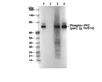 Lane 1: Ramos, Lane 2: Ramos ( λ phosphatase-treated), Lane 3: Mouse brain, Lane 4: Rat brain
Usage Information
| Dilution |
|---|
|
| Application |
|---|
| WB, IP |
| Reactivity |
|---|
| Human, Mouse, Rat |
| Source |
|---|
| Rabbit Monoclonal Antibody |
| Storage Buffer |
|---|
| PBS, pH 7.2+50% Glycerol+0.05% BSA+0.01% NaN3 |
| Storage (from the date of receipt) |
|---|
| -20°C (avoid freeze-thaw cycles), 2 years |
| Predicted MW |
|---|
| 78 kDa, 80 kDa, 82 kDa, 85 kDa |
| Positive Control | Mouse brain; Rat brain; OVCAR8 cell; Ramos cell |
|---|---|
| Negative Control | OVCAR8 (λ phosphatase-treated); Ramos (λ phosphatase-treated) |
Experimental Methods
| WB |
|---|
Experimental Protocol:
Sample preparation
1. Tissue: Lyse the tissue sample by adding an appropriate volume of ice-cold RIPA/NP-40 Lysis Buffer (containing Protease Inhibitor Cocktail, Phosphatase Inhibitor Cocktail),and homogenize the tissue at a low temperature. 2. Adherent cell: Aspirate the culture medium and wash the cells with ice-cold PBS twice. Lyse the cells by adding an appropriate volume of RIPA/NP-40 Lysis Buffer (containing Protease Inhibitor Cocktail, Phosphatase Inhibitor Cocktail) and put the sample on ice for 5 min. 3. Suspension cell: Transfer the culture medium to a pre-cooled centrifuge tube. Centrifuge and aspirate the supernatant. Wash the cells with ice-cold PBS twice. Lyse the cells by adding an appropriate volume of RIPA/NP-40 Lysis Buffer (containing Protease Inhibitor Cocktail, Phosphatase Inhibitor Cocktail) and put the sample on ice for 5 min. 4. Place the lysate into a pre-cooled microcentrifuge tube. Centrifuge at 4°C for 15 min. Collect the supernatant;
5. Remove a small volume of lysate to determine the protein concentration;
6. Combine the lysate with protein loading buffer. Boil 20 µL sample under 95-100°C for 5 min. Centrifuge for 5 min after cool down on ice.
Electrophoretic separation
1. According to the concentration of extracted protein, load appropriate amount of protein sample and marker onto SDS-PAGE gels for electrophoresis. Recommended separating gel (lower gel) concentration: 10%. Reference Table for Selecting SDS-PAGE Separation Gel Concentrations 2. Power up 80V for 30 minutes. Then the power supply is adjusted (110 V~150 V), the Marker is observed, and the electrophoresis can be stopped when the indicator band of the predyed protein Marker where the protein is located is properly separated. (Note that the current should not be too large when electrophoresis, too large current (more than 150 mA) will cause the temperature to rise, affecting the result of running glue. If high currents cannot be avoided, an ice bath can be used to cool the bath.)
Transfer membrane
1. Take out the converter, soak the clip and consumables in the pre-cooled converter;
2. Activate PVDF membrane with methanol for 1 min and rinse with transfer buffer;
3. Install it in the order of "black edge of clip - sponge - filter paper - filter paper - glue -PVDF membrane - filter paper - filter paper - sponge - white edge of clip"; 4. The protein was electrotransferred to PVDF membrane. ( 0.45 µm PVDF membrane is recommended ) Reference Table for Selecting PVDF Membrane Pore Size Specifications Recommended conditions for wet transfer: 200 mA, 120 min. ( Note that the transfer conditions can be adjusted according to the protein size. For high-molecular-weight proteins, a higher current and longer transfer time are recommended. However, ensure that the transfer tank remains at a low temperature to prevent gel melting.)
Block
1. After electrotransfer, wash the film with TBST at room temperature for 5 minutes;
2. Incubate the film in the blocking solution ( recommending 5% BSA solution)
for 1 hour at room temperature;
3. Wash the film with TBST for 3 times, 5 minutes each time.
Antibody incubation
1. Use 5% skim milk powder to prepare the primary antibody working liquid (recommended dilution ratio for primary antibody 1:1000), gently shake and incubate with the film at 4°C overnight; 2. Wash the film with TBST 3 times, 5 minutes each time;
3. Add the secondary antibody to the blocking solution and incubate with the film gently at room temperature for 1 hour;
4. After incubation, wash the film with TBST 3 times for 5 minutes each time.
Antibody staining
1. Add the prepared ECL luminescent substrate (or select other color developing substrate according to the second antibody) and mix evenly;
2. Incubate with the film for 1 minute, remove excess substrate (keep the film moist), wrap with plastic film, and expose in the imaging system. |
Biological Description
| Specificity |
|---|
Phospho-PKC (pan) (γ Thr514) Antibody [C12D18] recognizes endogenous levels of PKC α, β I, β II, γ, δ, ε, η, and θ isoforms only when phosphorylated at Thr 514. |
| Subcellular Location |
|---|
| Cytoplasm, Membrane, Nucleus |
| Uniprot ID |
|---|
| P05771, P17252, P24723, Q05655, Q04759, Q02156, P05129 |
| Clone |
|---|
| C12D18 |
| Synonym(s) |
|---|
| Protein kinase C beta type, PKC-B; PKC-beta, PRKCB, PKCB, PRKCB1, Protein kinase C alpha type, PKC-A; PKC-alpha, PRKCA, PKCA, PRKACA, Protein kinase C eta type, PKC-L, nPKC-eta, PRKCH, PKCL, PRKCL, Protein kinase C delta type, Tyrosine-protein kinase PRKCD, nPKC-delta, PRKCD, PKCD, Protein kinase C theta type, nPKC-theta, PRKCQ, PRKCT, Protein kina |
| Background |
|---|
| Phosphorylation of PKCγ at threonine 514 (Thr514) is a pivotal event in the maturation and regulation of this serine/threonine kinase, marking the first step in a sequence of phosphorylation events that prime the enzyme for activation. This phosphorylation, catalyzed by PDK-1, occurs within the activation loop of the kinase domain and is essential for stabilizing the structure of newly synthesized PKCγ in a catalytically competent yet inactive state. This allows PKCγ to be stored in the cytoplasm, ready to respond rapidly to cellular stimuli. Upon stimulation, typically involving elevated intracellular calcium and diacylglycerol, PKCγ undergoes conformational changes and translocates to the membrane, where it becomes fully activated and participates in signal transduction. Thr514 phosphorylation is thus a prerequisite for the enzyme’s proper subcellular localization, activation, and function. phospho-PKCγ plays critical roles in regulating cell proliferation, differentiation, migration, and survival, particularly in neurons and epithelial cells. Dysregulation at this site, such as aberrant phosphorylation or expression, results in cancer progression in tissues like the colon. |
| References |
|---|
|
Tech Support
Tel: +1-832-582-8158 Ext:3
If you have any other enquiries, please leave a message.
Products are for research use only. Not for human use. We do not sell to patients.
©Copyright 2013 Selleck Chemicals. All Rights Reserved.