-
 Australia
-
 Austria
-
 Belgium
-
 Brazil
-
 Canada
-
 China
China
-
 Czech Republic
-
 Denmark
-
 Finland
-
 France
-
 Germany
-
 Greece
-
 Hong Kong
-
 Hungary
-
 Iceland
-
 India
India
-
 Ireland
-
 Israel
-
 Italy
Italy
-
 Japan
Japan
-
 Korea
Korea
-
 Luxembourg
-
 Malaysia
-
 Netherlands
-
 New Zealand
-
 Norway
-
 Poland
-
 Qatar
Qatar
-
 Romania
-
 Saudi Arabia
-
 Singapore
-
 Spain
Spain
-
 Sweden
-
 Switzerland
-
 Taiwan
-
 Turkey
-
 United Kingdom
-
 United States
research use only
Phospho-β Catenin (Ser37) Antibody [H12E11]
Cat.No.: F2982
Application:
Reactivity:
-
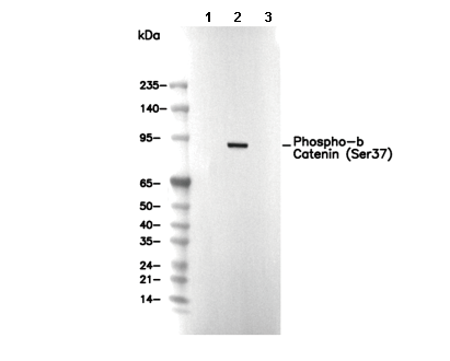 Lane 1: HEK-293, Lane 2: HEK-293 (Calyculin A, 50nM, 3 h), Lane 3: HEK-293 (Calyculin A, 50nM, 3 h; alkaline phosphatase, 1 h)
Customer Review
Usage Information
| Dilution |
|---|
|
| Application |
|---|
| WB |
| Reactivity |
|---|
| Human, Mouse, Rat |
| Source |
|---|
| Rabbit Monoclonal Antibody |
| Storage Buffer |
|---|
| PBS, pH 7.2+50% Glycerol+0.05% BSA+0.01% NaN3 |
| Storage (from the date of receipt) |
|---|
| -20°C (avoid freeze-thaw cycles), 2 years |
| Predicted MW |
|---|
| 85 kDa |
| Positive Control | C6 cell (treated with 100ng/ml Calyculin A for 30 min); NIH/3T3 cell (treated with 100nM Calyculin A for 30 min); HeLa cell (treated with 100nM Calyculin A for 30 min); HEK-293 cell (treated with 50nM Calyculin A for 3 hours) |
|---|---|
| Negative Control | C6 cell; NIH/3T3 cell; HeLa cell; HEK-293 cell |
Experimental Methods
| WB |
|---|
Experimental Protocol:
Sample preparation
1. Tissue: Lyse the tissue sample by adding an appropriate volume of ice-cold RIPA/NP-40 Lysis Buffer (containing Protease Inhibitor Cocktail, Phosphatase Inhibitor Cocktail),and homogenize the tissue at a low temperature. 2. Adherent cell: Aspirate the culture medium and wash the cells with ice-cold PBS twice. Lyse the cells by adding an appropriate volume of RIPA/NP-40 Lysis Buffer (containing Protease Inhibitor Cocktail, Phosphatase Inhibitor Cocktail) and put the sample on ice for 5 min. 3. Suspension cell: Transfer the culture medium to a pre-cooled centrifuge tube. Centrifuge and aspirate the supernatant. Wash the cells with ice-cold PBS twice. Lyse the cells by adding an appropriate volume of RIPA/NP-40 Lysis Buffer (containing Protease Inhibitor Cocktail, Phosphatase Inhibitor Cocktail) and put the sample on ice for 5 min. 4. Place the lysate into a pre-cooled microcentrifuge tube. Centrifuge at 4°C for 15 min. Collect the supernatant;
5. Remove a small volume of lysate to determine the protein concentration;
6. Combine the lysate with protein loading buffer. Boil 20 µL sample under 95-100°C for 5 min. Centrifuge for 5 min after cool down on ice.
Electrophoretic separation
1. According to the concentration of extracted protein, load appropriate amount of protein sample and marker onto SDS-PAGE gels for electrophoresis. Recommended separating gel (lower gel) concentration: 10%. Reference Table for Selecting SDS-PAGE Separation Gel Concentrations 2. Power up 80V for 30 minutes. Then the power supply is adjusted (110 V~150 V), the Marker is observed, and the electrophoresis can be stopped when the indicator band of the predyed protein Marker where the protein is located is properly separated. (Note that the current should not be too large when electrophoresis, too large current (more than 150 mA) will cause the temperature to rise, affecting the result of running glue. If high currents cannot be avoided, an ice bath can be used to cool the bath.)
Transfer membrane
1. Take out the converter, soak the clip and consumables in the pre-cooled converter;
2. Activate PVDF membrane with methanol for 1 min and rinse with transfer buffer;
3. Install it in the order of "black edge of clip - sponge - filter paper - filter paper - glue -PVDF membrane - filter paper - filter paper - sponge - white edge of clip"; 4. The protein was electrotransferred to PVDF membrane. ( 0.45 µm PVDF membrane is recommended ) Reference Table for Selecting PVDF Membrane Pore Size Specifications Recommended conditions for wet transfer: 200 mA, 120 min. ( Note that the transfer conditions can be adjusted according to the protein size. For high-molecular-weight proteins, a higher current and longer transfer time are recommended. However, ensure that the transfer tank remains at a low temperature to prevent gel melting.)
Block
1. After electrotransfer, wash the film with TBST at room temperature for 5 minutes;
2. Incubate the film in the blocking solution ( recommending 5% BSA solution)
for 1 hour at room temperature;
3. Wash the film with TBST for 3 times, 5 minutes each time.
Antibody incubation
1. Use 5% skim milk powder to prepare the primary antibody working liquid (recommended dilution ratio for primary antibody 1:1000), gently shake and incubate with the film at 4°C overnight; 2. Wash the film with TBST 3 times, 5 minutes each time;
3. Add the secondary antibody to the blocking solution and incubate with the film gently at room temperature for 1 hour;
4. After incubation, wash the film with TBST 3 times for 5 minutes each time.
Antibody staining
1. Add the prepared ECL luminescent substrate (or select other color developing substrate according to the second antibody) and mix evenly;
2. Incubate with the film for 1 minute, remove excess substrate (keep the film moist), wrap with plastic film, and expose in the imaging system. |
Biological Description
| Specificity |
|---|
Phospho-β Catenin (Ser37) Antibody [H12E11] detects endogenous levels of β-catenin only when phosphorylated at serines 37. |
| Subcellular Location |
|---|
| Cell junction, Cell membrane, Cell projection, Cytoplasm, Cytoskeleton, Membrane, Nucleus, Synapse |
| Uniprot ID |
|---|
| P35222 |
| Clone |
|---|
| H12E11 |
| Synonym(s) |
|---|
| CTNNB, OK/SW-cl.35, PRO2286, CTNNB1, Catenin beta-1, Beta-catenin |
| Background |
|---|
| Phospho-β Catenin (Ser37) is a multifunctional protein belonging to the armadillo family, characterized by 12 armadillo repeats forming a superhelical structure with a positively charged groove that serves as a versatile interface for binding partners. It is a 781-amino-acid-long protein consisting of the N-terminal domain (NTD), twelve armadillo (ARM) domains in the middle of the protein, and the C-terminal domain (CTD). It plays dual roles in cadherin-mediated cell-cell adhesion and Wnt signaling-mediated transcriptional regulation. In adhesion, β-catenin stabilizes cadherins at the membrane and recruits α-catenin to link to the actin cytoskeleton, while in signaling, it acts as a co-activator in the LEF/TCF transcription complex upon Wnt activation. The function of β-catenin is tightly regulated by its phosphorylation status; phosphorylation at Ser37 (along with S33 and T41) by GSK3β or PKC targets β-catenin for ubiquitination and proteasomal degradation, thus preventing aberrant Wnt signaling. Ligand phosphorylation can also enhance β-catenin’s binding affinity by increasing negative charges that interact with its armadillo domain. Its unstructured N- and C-terminal regions further modulate interaction specificity and accessibility. Altogether, β-catenin’s structural adaptability and phosphorylation-dependent regulation are critical for its dynamic roles in tissue morphogenesis, cell fate decisions, and maintenance of tissue integrity. |
| References |
|---|
|
Tech Support
Tel: +1-832-582-8158 Ext:3
If you have any other enquiries, please leave a message.
Products are for research use only. Not for human use. We do not sell to patients.
©Copyright 2013 Selleck Chemicals. All Rights Reserved.