-
 Australia
-
 Austria
-
 Belgium
-
 Brazil
-
 Canada
-
 China
China
-
 Czech Republic
-
 Denmark
-
 Finland
-
 France
-
 Germany
-
 Greece
-
 Hong Kong
-
 Hungary
-
 Iceland
-
 India
India
-
 Ireland
-
 Israel
-
 Italy
Italy
-
 Japan
Japan
-
 Korea
Korea
-
 Luxembourg
-
 Malaysia
-
 Netherlands
-
 New Zealand
-
 Norway
-
 Poland
-
 Qatar
Qatar
-
 Romania
-
 Saudi Arabia
-
 Singapore
-
 Spain
Spain
-
 Sweden
-
 Switzerland
-
 Taiwan
-
 Turkey
-
 United Kingdom
-
 United States
research use only
Pelitinib (EKB-569) EGFR inhibitor
Cat.No.S1392
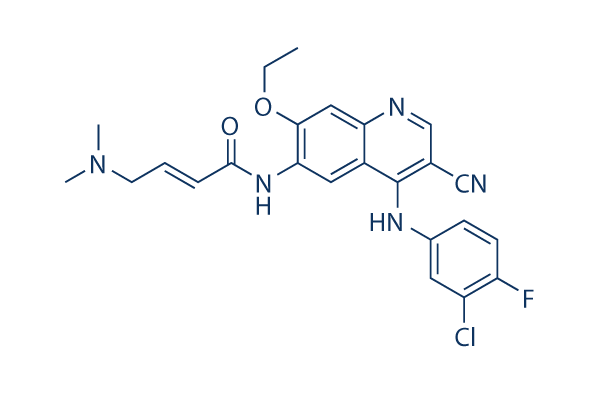
Chemical Structure
Molecular Weight: 467.92
Quality Control
| Related Targets | VEGFR PDGFR FGFR c-Met Src MEK CSF-1R FLT3 HER2 c-Kit |
|---|---|
| Other EGFR Inhibitors | Lazertinib (YH25448) Icotinib Hydrochloride Sunvozertinib AG-490 AG-1478 Canertinib (CI-1033) WZ4002 Poziotinib (NOV120101, HM781-36B) Rociletinib (CO-1686) Genistein |
Cell Culture, Treatment & Working Concentration
| Cell Lines | Assay Type | Concentration | Incubation Time | Formulation | Activity Description | PMID |
|---|---|---|---|---|---|---|
| A431 | Function assay | Inhibition of EGFR autophosphorylation in human A431 cells, IC50=0.00802μM. | 20797871 | |||
| Sf9 | Function assay | 10 mins | Inhibition of recombinant human His6-tagged EGFR cytoplasmic domain (645 to 1186 residues) expressed in baculovirus infected Sf9 insect cells assessed as reduction in autophosphorylation preincubated for 10 mins followed by ATP-MgCl2 addition and measured, IC50=0.0385μM. | 30600149 | ||
| A431 | Function assay | 90 mins | Inhibition of EGFR in human A431 cells assessed as reduction in EGF-stimulated EGFR autophosphorylation preincuabted for 90 mins followed by EGF-stimulation by sandwich-ELISA, IC50=0.039μM. | 30973735 | ||
| DiFi | Function assay | 2 hrs | Inhibition autophosphorylation of EGFR in human DiFi cells after 2 hrs by ELISA, IC50=0.07943μM. | 17689836 | ||
| UCH1 | Antiproliferative assay | 72 hrs | Antiproliferative activity against human UCH1 cells measured after 72 hrs by alamar blue assay, IC50=0.09μM. | 30973735 | ||
| UCH2 | Antiproliferative assay | 72 hrs | Antiproliferative activity against human UCH2 cells measured after 72 hrs by alamar blue assay, IC50=1.6μM. | 30973735 | ||
| Click to View More Cell Line Experimental Data | ||||||
Solubility
|
In vitro |
DMSO
: 13 mg/mL
(27.78 mM)
Water : Insoluble Ethanol : Insoluble |
Molarity Calculator
|
In vivo |
|||||
In vivo Formulation Calculator (Clear solution)
Step 1: Enter information below (Recommended: An additional animal making an allowance for loss during the experiment)
Step 2: Enter the in vivo formulation (This is only the calculator, not formulation. Please contact us first if there is no in vivo formulation at the solubility Section.)
Calculation results:
Working concentration: mg/ml;
Method for preparing DMSO master liquid: mg drug pre-dissolved in μL DMSO ( Master liquid concentration mg/mL, Please contact us first if the concentration exceeds the DMSO solubility of the batch of drug. )
Method for preparing in vivo formulation: Take μL DMSO master liquid, next addμL PEG300, mix and clarify, next addμL Tween 80, mix and clarify, next add μL ddH2O, mix and clarify.
Method for preparing in vivo formulation: Take μL DMSO master liquid, next add μL Corn oil, mix and clarify.
Note: 1. Please make sure the liquid is clear before adding the next solvent.
2. Be sure to add the solvent(s) in order. You must ensure that the solution obtained, in the previous addition, is a clear solution before proceeding to add the next solvent. Physical methods such
as vortex, ultrasound or hot water bath can be used to aid dissolving.
Chemical Information, Storage & Stability
| Molecular Weight | 467.92 | Formula | C24H23ClFN5O2 |
Storage (From the date of receipt) | |
|---|---|---|---|---|---|
| CAS No. | 257933-82-7 | Download SDF | Storage of Stock Solutions |
|
|
| Synonyms | N/A | Smiles | CCOC1=C(C=C2C(=C1)N=CC(=C2NC3=CC(=C(C=C3)F)Cl)C#N)NC(=O)C=CCN(C)C | ||
Mechanism of Action
| Features |
An improved version of EKI-785.
|
|---|---|
| Targets/IC50/Ki |
EGFR
38.5 nM
Src
282 nM
MEK/ERK
800 nM
ErbB2
1.255 μM
Raf
3.353 μM
|
| In vitro |
Pelitinib (EKB-569) displays much higher inhibitory activity against EGFR, compared with the closely related c-erbB-2, as well as other kinases such as Src, Cdk4, c-Met, Raf, and MEK/ERK, with IC50 ranging from 282 nM for Src to >20 μM for Cdk4. Consistently, this compound treatment significantly inhibits the autophosphorylation of EGFR but not c-Met in A431 cells. It potently inhibits the proliferation of normal human keratinocytes (NHEK), as well as A431 and MDA-468 tumor cells with IC50 of 61 nM, 125 nM, and 260 nM, respectively, while displaying little activity against MCF-7 cells with IC50 of 3.6 μM. This chemical inhibits EGF-induced phosphorylation of EGFR in A431 and NHEK cells with IC50 of 20-80 nM, as well as the phosphorylation of STAT3 with IC50 of 30-70 nM. It at 75-500 nM also specifically inhibits the activation of AKT and ERK1/2, without affecting NF-κB pathway. In NHEK cells, this compound also potently inhibits TGF-α mediated EGFR activation with IC50 of 56 nM, as well as activation of STAT3 and ERK1/2 with IC50 of 60 nM and 62 nM, respectively.
|
| Kinase Assay |
Autophosphorylation of EGFR in cells
|
|
For experiments using cells in culture, A431 cells are treated with various concentrations of Pelitinib (EKB-569) for 2.75 hours before co-incubation with 100 ng/mL EGF for 0.25 hour. Cells are washed twice with cold phosphate-buffered saline (PBS) before adding to lysis buffer (10 mM Tris, pH 7.5, 5 mM ethylenediamine tetra-acetic acid (EDTA), 150 mM NaCl, 1% Triton X-100, 1% Sodium deoxycholate, 0.1 % SDS, 1 mM PMSF, 10 mg/mL pepstatin A, 10 mg/mL leupeptin, 20 KIU/mL aprotinin, 2 mM sodium orthovanadate, and 100 mM sodium fluoride) for 20 minutes on ice, before immunoprecipitation and SDS-PAGE-immunoblotting. For immunoprecipitation, cultured cells are placed in cold lysis buffer and immediately homogenized on ice with a polytron with several pulses. The homogenate is first centrifuged at 2500 rpm (20 minutes, 4 °C) and then again at 14,000 rpm in a microcentrifuge (10 minutes, 4 °C). Supernatants (1000 μg protein) are incubated for 2 hours at 4 °C with 15 mL of EGFR polyclonal antibody. After 2 hours, 50 μL of protein G plus/protein A agarose beads is added and incubated with constant rotation for 2 hours at 4 °C. After washing with lysis buffer, beads are boiled for 2 minutes in Laemmli sample buffer. Proteins are then resolved by SDS-PAGE, transferred to immobilon membrane and probed overnight with an anti-phosphotyrosine antibody conjugated with horseradish peroxidase (HRP). Membranes are developed using the ECL reagent. Total EGFR protein is determined by stripping membranes and re-probing with receptor-specific antibodies. Quantitation of bands is done by densitometry, using ImageQuant software with a Molecular Dynamics laser transmittance scanner.
|
|
| In vivo |
A single oral dose of 10 mg/kg Pelitinib (EKB-569) potently inhibits the EGFR phosphorylation in A431 xenografts with over-expressed EGFR, by 90% within 1 hour, and by >50% after 24 hours. Administration of this compound at 20 mg/kg/day inhibits tumorigenesis in APCMin/+ mice by 87%, equivalent to the effect of used with 2 times doses of EKI-785 (40 mg/kg/day), consistent with greater in vivo potency. This chemical selectively inhibits EGFR signaling in airway epithelial cells in vivo. In the mouse model of airway epithelial remodeling that is inducible by viral infection and features a delayed but permanent switch to goblet cell metaplasia, this compound treatment at 20 mg/kg/day corrects all 3 aspects of epithelial remodeling, by completely blocking the increase of ciliated cells and decrease of Clara cells, and significantly inhibiting the metaplasia of goblet cells.
|
References |
|
Tech Support
Tel: +1-832-582-8158 Ext:3
If you have any other enquiries, please leave a message.
Signaling Pathway Map
Products are for research use only. Not for human use. We do not sell to patients.
©Copyright 2013 Selleck Chemicals. All Rights Reserved.