
Why did 10% FBS reduce SCH772984 efficacy against downstream p-Elk1 compared to serum-free conditions?
Quick Answer
Recommended ApproachSerum-starve cells prior to inhibitor treatment when evaluating downstream p-Elk1 suppression, or adjust compound concentrations upward to account for serum protein binding and mitogenic drive.
Common CausesTesting SCH772984 in 10% FBS reduced downstream Elk-1 dephosphorylation because serum albumin sequestered the lipophilic compound while serum growth factors intensely stimulated upstream RTK-MEK signaling flux.
How Should It Be Done Correctly?
Culture and Dosing Matrix for MAPK Substrate Assays
When evaluating target engagement and downstream substrate phosphorylation (such as phospho-Elk1) by the ERK1/2 inhibitor SCH772984, the assay medium composition dictates the effective free drug fraction and baseline kinase flux [1, 2].
| Experimental Parameter | Serum-Starved Condition (Recommended for MoA) | Complete Medium Condition (10% FBS) |
|---|---|---|
| Primary Objective | Direct target engagement, intrinsic biochemical potency | Long-term viability, proliferation, chronic signaling [1] |
| Pre-treatment Starvation | 4 to 16 hours in 0% to 0.1% BSA medium | None (maintained in growth medium with 10% FBS) |
| Free Drug Availability | High (>95% unbound compound) | Low (~10% to 20% unbound due to albumin binding) |
| Upstream RTK-MEK Drive | Basal / quiescent | Sustained, high mitogenic signaling flux |
| Expected IC50 Shift (p-Elk1) | Baseline reference (low nanomolar) | 5- to 10-fold rightward shift (higher concentration needed) |
| Vehicle (DMSO) Limit | ≤ 0.1% (v/v) final in culture | ≤ 0.1% (v/v) final in culture |
| Compound Exposure Window | 1 to 2 hours (acute) up to 24 hours [1, 3] | 24 to 72 hours (chronic phenotypic readouts) [1] |
Step-by-Step Procedure for Downstream Substrate Readouts
- Plate Preparation and Starvation: Seed target cells (such as melanoma or NSCLC lines) in standard multi-well plates and culture until they reach approximately 70% confluence [1, 3]. Aspirate growth medium, wash twice with pre-warmed PBS to clear residual bovine serum components, and replace with serum-free basal medium (or basal medium supplemented with 0.1% fatty-acid-free BSA) for 4 to 16 hours to synchronize cells and silence exogenous RTK stimulation.
- Compound Dilution and Preparation: Reconstitute SCH772984 stock in high-quality anhydrous DMSO (up to 14 mg/mL according to solubility specifications). Prepare an intermediate working dilution in serum-free medium immediately prior to use. Ensure final in-well DMSO concentrations do not exceed 0.1% (v/v) to avoid vehicle-induced membrane permeabilization or non-specific signaling shifts.
- Inhibitor Pre-Incubation: Apply serial dilutions of SCH772984 directly to the serum-starved cells. Pre-incubate for 30 to 60 minutes at 37°C in a humidified 5% CO2 incubator to allow complete intracellular accumulation and binding to ERK1/2 [1, 2].
- Mitogen Challenge (Optional Dynamic Assays): If assessing the ability of SCH772984 to block mitogen-induced Elk-1 activation, pulse the pre-treated cells with an acute stimulus (such as 10% FBS or 50 ng/mL recombinant EGF) for 15 to 30 minutes. If evaluating steady-state basal signaling, omit the pulse.
- Rapid Cell Lysis and Quenching: Immediately aspirate the medium, place plates on ice, and rinse once with ice-cold PBS. Lyse cells rapidly in RIPA or urea-based lysis buffer containing freshly added protease inhibitors, sodium orthovanadate, and sodium fluoride to arrest all kinase and phosphatase activity instantaneously.
- Phosphoprotein Immunoblotting: Resolve normalized protein lysates via SDS-PAGE and transfer to nitrocellulose or PVDF membranes. Probe with validated antibodies against phospho-Elk1 (Ser383), total Elk-1, phospho-ERK1/2 (Thr202/Tyr204), and total ERK1/2, utilizing an invariant housekeeping protein (such as beta-actin) as a loading standard [1, 2].
What Went Wrong?
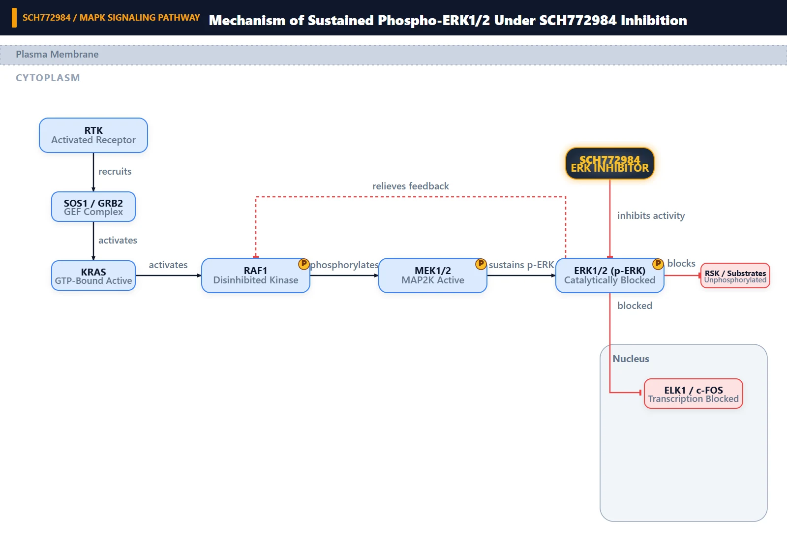
Copy code to add this image to your page Copy Code
Testing Intrinsic Downstream Potency in High-Serum Formulations
The investigator assayed SCH772984-mediated inhibition of downstream Elk-1 phosphorylation in culture medium containing 10% fetal bovine serum (FBS) and directly compared the resulting potency against serum-free conditions. Testing competitive kinase inhibitors in full serum for acute downstream biochemical readouts without compensating for serum components introduces major confounding variables that degrade observed inhibitory efficacy.
Non-Specific Serum Sequestration and Mitogen-Driven Signal Amplification
The proximate cause of the reduced efficacy is twofold: extensive non-specific binding of SCH772984 to bovine serum proteins, and persistent mitogenic stimulation supplied by FBS. Fetal bovine serum is rich in serum albumin and alpha-1-acid glycoprotein. Hydrophobic small molecules like SCH772984 partition into serum albumin, substantially reducing the free (unbound) fraction available to traverse the plasma membrane and engage ERK1/2 inside the cell. Concurrently, growth factors in 10% FBS activate membrane-bound receptor tyrosine kinases (RTKs), sustaining continuous catalytic flux through the pathway and requiring higher total drug levels to achieve equal substrate dephosphorylation.
High Upstream Catalytic Flux and Feedback Disinhibition
At the molecular level, Elk-1 is a direct nuclear target of ERK1/2, phosphorylated specifically at Ser383 to drive immediate-early gene transcription. SCH772984 functions as an ATP-competitive inhibitor of ERK1 and ERK2 [1, 2].
Under serum-starved conditions, basal signaling through the RAS-RAF-MEK cascade is minimal, meaning intracellular ERK1/2 maintains low baseline turnover; a modest concentration of SCH772984 is sufficient to saturate active sites and extinguish residual Elk-1 phosphorylation. In contrast, 10% FBS supplies abundant ligands (EGF, FGF, PDGF, IGF-1) that trigger RTK dimerization and intense activation of RAS and RAF, driving hyperactivation of MEK1/2 [1, 2]. MEK1/2 continuously phosphorylates the activation loop (Thr202/Tyr204) of ERK1/2 [2].
Furthermore, ERK inhibition relieves upstream negative feedback loops (such as the inhibitory phosphorylation of RAF1 at Ser43 and son of sevenless [SOS]), resulting in compensatory MEK hyperactivation [2]. When upstream MEK phosphorylation of ERK is elevated, active ERK enzymes outcompete the inhibitor or saturate nuclear substrate pools, resulting in a pronounced rightward shift in the IC50 for phospho-Elk1.
How Can I Avoid This in Future Experiments?
To prevent serum-dependent discrepancy in future target validation assays, explicitly decouple intrinsic biochemical target engagement from phenotypic proliferation assays. Use short-term serum starvation (4–16 hours) whenever determining IC50 values for downstream substrate dephosphorylation (e.g., p-Elk1, p-RSK), which minimizes mitogen noise and eliminates protein-binding artifacts [1, 2]. If phenotypic responses must be characterized in complete 10% FBS (such as 72-hour viability or colony formation assays), run a parallel serum-shift binding assay to quantify the free drug fraction and calibrate the working concentration accordingly.