
Why does p-ERK1/2 paradoxical hyperactivation persist after 1 µM SCH772984 treatment?
Quick Answer
Recommended ApproachMonitor functional downstream substrates such as phospho-p90RSK rather than phospho-ERK1/2 alone, and co-target upstream nodes like MEK or RAF to suppress feedback-driven pathway rebound.
Common CausesInterpreting elevated phospho-ERK1/2 as active signaling fails because catalytic ERK inhibition relieves negative feedback on RAF1 and MEK, causing upstream hyperactivation and accumulation of phosphorylated ERK.
How Should It Be Done Correctly?
Analytical and Dosing Parameters for MAPK Target Validation
To accurately evaluate ERK1/2 target engagement without being misled by feedback-induced phosphosite accumulation, the assay design must account for dynamic temporal rebounds, proper readouts, and combined upstream pathway suppression.
| Parameter | Recommended Specification | Experimental Role / Threshold |
|---|---|---|
| Primary Readout Target | Phospho-p90RSK (p-RSK Ser380) | Bona fide downstream functional output of active ERK1/2 [1] |
| Secondary Readout Target | Phospho-MEK1/2 (p-MEK Ser217/221) | Diagnostic indicator of upstream pathway feedback relief [1, 2] |
| Reference Readout Target | Phospho-ERK1/2 (Thr202/Tyr204) | Physical target engagement/phosphorylation status only [1] |
| Compound Stock Solvent | Anhydrous DMSO (≤ 14 mg/mL) | Stock preparation at -20°C (1 mo) or -80°C (1 yr) |
| In Vitro Working Dose | 250 nM – 1 µM | Target engagement threshold depending on baseline sensitivity [1] |
| Dynamic Assessment Times | 1 h (acute), 6 h, 24 h, 48 h (extended) | Captures initial pathway shutdown versus adaptive rebound [1] |
| Feedback Blockade Strategy | SCH772984 + Trametinib / Vemurafenib | Dual vertical inhibition preventing MEK/ERK feedback rebound [1] |
Operational Procedure for Dynamic Kinase Pathway Profiling
-
Stock Preparation and Cell Seeding - Dissolve solid SCH772984 in anhydrous DMSO at a concentration not exceeding 14 mg/mL according to product solubility limits. Store single-use aliquots at -80°C. - Seed cells (e.g., BRAF- or NRAS-mutant melanoma or KRAS-driven carcinoma lines) in multi-well culture plates at optimal density to reach 70–80% confluence at the experimental endpoint [1, 3]. Allow cells to adhere and equilibrate overnight in complete medium.
-
Compound Dilution and Treatment Execution - Prepare intermediate dilutions in fresh, pre-warmed culture media immediately prior to dosing; ensure the final vehicle DMSO concentration remains ≤ 0.1% (v/v) across all wells [Source: r/labrats]. - Apply SCH772984 across a standardized multi-point time course: include 1 h, 2 h, 6 h, 12 h, 24 h, and 48 h post-treatment cohorts alongside matched DMSO vehicle controls [1]. For combination controls, incorporate parallel cohorts receiving MEK inhibitor (e.g., trametinib) or RAF inhibitor (e.g., vemurafenib or dabrafenib) [1, 2].
-
Rapid Protein Extraction and Phosphatase Preservation - Terminate signaling at the exact target time point by immediately placing culture plates on wet ice and aspirating media. Wash monolayers once with ice-cold PBS. - Lyse cells directly on the plate with chilled RIPA buffer fortified with fresh protease and phosphatase inhibitor cocktails (containing sodium orthovanadate, sodium fluoride, and beta-glycerophosphate). - Clarify lysates by centrifugation at ≥ 14,000 × g at 4°C for 15 minutes, determine protein concentrations using a standard bicinchoninic acid (BCA) assay, and boil lysates in SDS-PAGE loading buffer.
-
Multiplexed Immunoblot Assessment and Interpretation - Resolve equal protein masses (20–30 µg/lane) across SDS-PAGE gels. Transfer to PVDF or nitrocellulose membranes. - Probe simultaneously for phospho-p90RSK, total RSK, phospho-ERK1/2 (Thr202/Tyr204), total ERK1/2, phospho-MEK1/2 (Ser217/221), total MEK, and loading controls (beta-actin or GAPDH) [1]. - Decision Point: If p-RSK is completely abolished at 1–2 h, SCH772984 has successfully engaged and inhibited ERK1/2 [1]. If p-ERK1/2 persists or increases at 12–24 h while p-MEK is sharply upregulated, verify that p-RSK remains suppressed; this confirms feedback relief rather than compound inefficacy or resistance [1, 2].
What Went Wrong?
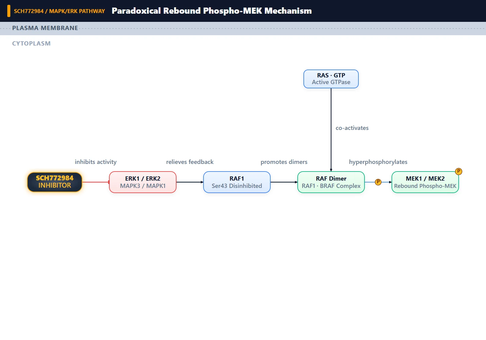
Copy code to add this image to your page Copy Code
The Incorrect Practice: Relying on Phospho-ERK1/2 as the Sole Inhibitory Marker
The misinterpretation arises from evaluating ERK inhibitor efficacy exclusively by measuring phospho-ERK1/2 (Thr202/Tyr204) band density at a single time point (e.g., 24 h post-treatment) without examining downstream substrate phosphorylation (p-RSK) or upstream activation states (p-MEK) [1]. Observing strong or increased phospho-ERK1/2 signals at 1 µM SCH772984 often leads researchers to prematurely conclude that the compound failed, degraded, or stimulated the pathway [1].
Proximate Cause: Rapid Relief of Inhibitory Feedback Loops
Under baseline conditions, active ERK1/2 phosphorylates several upstream regulatory nodes to dampen ongoing pathway flux [2]. When SCH772984 binds the ATP pocket of ERK1/2 and suppresses its catalytic activity, downstream effector signaling (such as p-RSK) drops precipitously within 1 hour [1]. However, this loss of ERK catalytic activity simultaneously strips the pathway of its negative feedback brakes [1, 2]. As a consequence, upstream kinase activity surges: RAF dimers and MEK1/2 undergo robust activation, yielding a massive accumulation of phosphorylated MEK (p-MEK Ser217/221) [1, 2]. Hyperactivated MEK continuously phosphorylates the Thr-Glu-Tyr (TEY) activation lip of ERK1/2, overwhelming basal cellular phosphatase activity and causing persistent, elevated p-ERK levels despite the catalytic site of ERK remaining drug-bound [1, 2].
Underlying Mechanism: RAF1 Dephosphorylation and Kinase Transphosphorylation Dynamics
The molecular mechanism driving paradoxical p-ERK1/2 elevation involves two distinct physical phenomena:
-
Feedback Dephosphorylation of RAF1 Ser43: ERK1/2 normally maintains direct negative feedback phosphorylation on upstream RAF kinases, notably at Ser43 on RAF1 (C-RAF) and homologous inhibitory sites on BRAF and SOS1 [2]. Upon pharmacological ERK inhibition by SCH772984, phosphorylation at RAF1 Ser43 is rapidly lost [2]. The removal of this inhibitory mark unleashes unrestrained RAF kinase activity, triggering downstream transphosphorylation of MEK1/2 at Ser217/Ser221 [1, 2]. Activated MEK then vigorously phosphorylates the activation loop (Thr202/Tyr204) of ERK1/2 [1].
-
Conformational Trapping and Catalytic Blockade: SCH772984 is a potent ATP-competitive ERK1/2 inhibitor [1, 3]. Although SCH772984 occupies the catalytic pocket and prevents ERK from phosphorylating its substrates (such as p90RSK, Elk-1, or c-Fos), it does not prevent upstream MEK from recognizing and phosphorylating the exposed activation loop of ERK [1]. In fact, occupancy of the ATP-binding site can sterically lock ERK1/2 into a conformation that shields Thr202/Tyr204 from dual-specificity phosphatases (DUSPs), while leaving the TEY motif fully accessible to the surging pool of hyperactivated MEK [1, 2]. Thus, the cell exhibits high steady-state p-ERK1/2 concomitant with marked p-MEK induction, while downstream pathway output remains suppressed or rebounds depending on the cell's genetic rewiring [1].
How Can I Avoid This in Future Experiments?
To avoid misinterpreting paradoxical activation in future studies, always run a vertical signaling panel that couples an ERK-specific downstream substrate (phospho-RSK Ser380) with upstream activation markers (phospho-MEK Ser217/221 and phospho-RAF) alongside phospho-ERK1/2 [1, 2]. A true loss of compound activity produces simultaneous recovery of both p-RSK and p-ERK without hyper-induced p-MEK, whereas feedback-driven paradoxical hyperactivation produces elevated p-ERK accompanied by supramaximal p-MEK induction while p-RSK remains suppressed [1].
Additionally, where sustained pathway shutdown is desired across cell line models that exhibit rapid feedback rebound, design combination protocols pairing SCH772984 with an upstream MEK inhibitor (e.g., trametinib) or RAF inhibitor (e.g., vemurafenib or dabrafenib) [1, 2]. Co-targeting MEK blocks the upstream feedback surge and prevents the accumulation of paradoxical phospho-ERK, establishing complete and durable MAPK pathway suppression [1].