-
 Australia
-
 Austria
-
 Belgium
-
 Brazil
-
 Canada
-
 China
China
-
 Czech Republic
-
 Denmark
-
 Finland
-
 France
-
 Germany
-
 Greece
-
 Hong Kong
-
 Hungary
-
 Iceland
-
 India
India
-
 Ireland
-
 Israel
-
 Italy
Italy
-
 Japan
Japan
-
 Korea
Korea
-
 Luxembourg
-
 Malaysia
-
 Netherlands
-
 New Zealand
-
 Norway
-
 Poland
-
 Qatar
Qatar
-
 Romania
-
 Saudi Arabia
-
 Singapore
-
 Spain
Spain
-
 Sweden
-
 Switzerland
-
 Taiwan
-
 Turkey
-
 United Kingdom
-
 United States
research use only
UQCRFS1/RISP Antibody [P24M17]
Cat.No.: F6358
Application:
Reactivity:
-
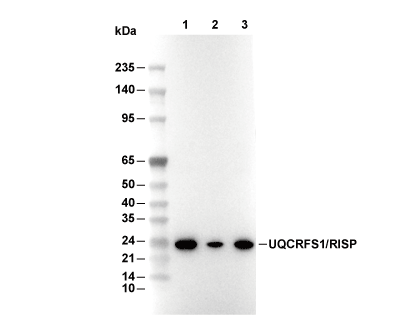 Lane 1: Neuro-2a, Lane 2: HT55, Lane 3: REC-1
Experiment Essentials
WB
Recommended wet transfer conditions: 200 mA, 60 min.
Recommended wet transfer conditions: 200 mA, 60 min.
Usage Information
| Dilution |
|---|
|
| Application |
|---|
| WB, IP |
| Reactivity |
|---|
| Human, Mouse, Rat |
| Source |
|---|
| Rabbit Monoclonal Antibody |
| Storage Buffer |
|---|
| PBS, pH 7.2+50% Glycerol+0.05% BSA+0.01% NaN3 |
| Storage (from the date of receipt) |
|---|
| -20°C (avoid freeze-thaw cycles), 2 years |
| Predicted MW |
|---|
| 23 kDa |
| Positive Control | HT55 cells; REC‑1 cells; HCC1806 cells; LOX‑IMVI cells; mIMCD‑3 cells; Neuro‑2a cells; NBT‑11 cells |
|---|---|
| Negative Control |
Experimental Methods
| WB |
|---|
Experimental Protocol:
Sample preparation
1. Tissue: Lyse the tissue sample by adding an appropriate volume of ice-cold RIPA/NP-40 Lysis Buffer (containing Protease Inhibitor Cocktail),and homogenize the tissue at a low temperature or lyse it by sonication on ice, then incubate on ice for 30 minutes. 2. Adherent cell: Aspirate the culture medium and wash the cells with ice-cold PBS twice. Lyse the cells by adding an appropriate volume of RIPA/NP-40 Lysis Buffer (containing Protease Inhibitor Cocktail), sonicate to lyse the cells, and incubate on ice for 30 minutes. 3. Suspension cell: Transfer the culture medium to a pre-cooled centrifuge tube. Centrifuge and aspirate the supernatant. Wash the cells with ice-cold PBS twice. Lyse the cells by adding an appropriate volume of RIPA/NP-40 Lysis Buffer (containing Protease Inhibitor Cocktail), sonicate to lyse the cells, and incubate on ice for 30 minutes. 4. Place the lysate into a pre-cooled microcentrifuge tube. Centrifuge at 4°C for 15 min. Collect the supernatant;
5. Remove a small volume of lysate to determine the protein concentration;
6. Combine the lysate with protein loading buffer. Boil 20 µL sample under 95-100°C for 5 min. Centrifuge for 5 min after cool down on ice.
Electrophoretic separation
1. According to the concentration of extracted protein, load appropriate amount of protein sample and marker onto SDS-PAGE gels for electrophoresis. Recommended separating gel (lower gel) concentration: 10%. Reference Table for Selecting SDS-PAGE Separation Gel Concentrations 2. Power up 80V for 30 minutes. Then the power supply is adjusted (110 V~150 V), the Marker is observed, and the electrophoresis can be stopped when the indicator band of the predyed protein Marker where the protein is located is properly separated. (Note that the current should not be too large when electrophoresis, too large current (more than 150 mA) will cause the temperature to rise, affecting the result of running glue. If high currents cannot be avoided, an ice bath can be used to cool the bath.)
Transfer membrane
1. Take out the converter, soak the clip and consumables in the pre-cooled converter;
2. Activate PVDF membrane with methanol for 1 min and rinse with transfer buffer;
3. Install it in the order of "black edge of clip - sponge - filter paper - filter paper - glue -PVDF membrane - filter paper - filter paper - sponge - white edge of clip"; 4. The protein was electrotransferred to PVDF membrane. ( 0.45 µm PVDF membrane is recommended ) Reference Table for Selecting PVDF Membrane Pore Size Specifications Recommended conditions for wet transfer: 200 mA, 60 min. ( Note that the transfer conditions can be adjusted according to the protein size. For high-molecular-weight proteins, a higher current and longer transfer time are recommended. However, ensure that the transfer tank remains at a low temperature to prevent gel melting.)
Block
1. After electrotransfer, wash the film with TBST at room temperature for 5 minutes;
2. Incubate the film in the blocking solution for 1 hour at room temperature;
3. Wash the film with TBST for 3 times, 5 minutes each time.
Antibody incubation
1. Use 5% skim milk powder to prepare the primary antibody working liquid (recommended dilution ratio for primary antibody 1:1000), gently shake and incubate with the film at 4°C overnight; 2. Wash the film with TBST 3 times, 5 minutes each time;
3. Add the secondary antibody to the blocking solution and incubate with the film gently at room temperature for 1 hour;
4. After incubation, wash the film with TBST 3 times for 5 minutes each time.
Antibody staining
1. Add the prepared ECL luminescent substrate (or select other color developing substrate according to the second antibody) and mix evenly;
2. Incubate with the film for 1 minute, remove excess substrate (keep the film moist), wrap with plastic film, and expose in the imaging system. |
Biological Description
| Specificity |
|---|
| UQCRFS1/RISP Antibody [P24M17] detects endogenous levels of total UQCRFS1/RISP protein. |
| Subcellular Location |
|---|
| Membrane, Mitochondrion, Mitochondrion inner membrane |
| Uniprot ID |
|---|
| P47985 |
| Clone |
|---|
| P24M17 |
| Synonym(s) |
|---|
| Cytochrome b-c1 complex subunit 5; Rieske iron-sulfur protein; RISP; UQCRFS1 |
| Background |
|---|
| UQCRFS1, known as the Rieske iron-sulfur protein or RISP, functions as the mobile extrinsic subunit of mitochondrial complex III (cytochrome bc1 complex) within the electron transport chain, facilitating electron shuttling from ubiquinol to cytochrome c during oxidative phosphorylation. The protein harbors a [2Fe-2S] cluster coordinated by eight cysteine and histidine residues within a folded extrinsic domain tethered by a single transmembrane helix, enabling conformational swinging between Qo and Qi sites of cytochrome b to execute the Q-cycle that translocates four protons per two electrons while preventing superoxide leakage under physiological potentials. During catalysis, reduced ubiquinol at the Qo site donates the first electron to the high-potential [2Fe-2S] cluster, which relays it to cytochrome c1 via histidine ligation, while the semiquinone intermediate bifurcates the second electron to bL and bH hemes, sustaining chain continuity and protonmotive force generation coupled to ATP synthesis. BCS1L chaperone imports the mitochondrial targeting sequence-cleaved precursor from the matrix, stabilizing the holoform through LYRM7-mediated Fe-S cluster insertion before membrane integration proximal to UQCRC1/UQCRC2 core subunits. In hematopoietic stem cells, UQCRFS1 deficiency disrupts quiescence by impairing TGF-β signaling coordination through SHP-1 interactions, elevating cycling indices and exhausting long-term repopulation capacity during serial transplantation. T cell receptor stimulation recruits UQCRFS1 to lipid rafts where complex III activity amplifies NFAT and NF-κB activation via localized ROS and calcium flux, with knockout diminishing IL-2 production and proliferation. |
| References |
|---|
|
Tech Support
Tel: +1-832-582-8158 Ext:3
If you have any other enquiries, please leave a message.
Products are for research use only. Not for human use. We do not sell to patients.
©Copyright 2013 Selleck Chemicals. All Rights Reserved.