-
 Australia
-
 Austria
-
 Belgium
-
 Brazil
-
 Canada
-
 China
China
-
 Czech Republic
-
 Denmark
-
 Finland
-
 France
-
 Germany
-
 Greece
-
 Hong Kong
-
 Hungary
-
 Iceland
-
 India
India
-
 Ireland
-
 Israel
-
 Italy
Italy
-
 Japan
Japan
-
 Korea
Korea
-
 Luxembourg
-
 Malaysia
-
 Netherlands
-
 New Zealand
-
 Norway
-
 Poland
-
 Qatar
Qatar
-
 Romania
-
 Saudi Arabia
-
 Singapore
-
 Spain
Spain
-
 Sweden
-
 Switzerland
-
 Taiwan
-
 Turkey
-
 United Kingdom
-
 United States
research use only
DUSP6/MKP3 Antibody [E9K23]
Cat.No.: F8982
Application:
Reactivity:
-
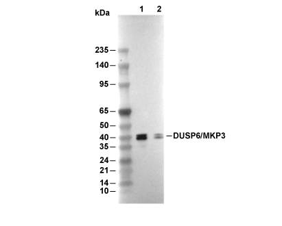 Lane 1: NCI-H3255, Lane 2: DLD-1
Usage Information
| Dilution |
|---|
|
| Application |
|---|
| WB, IP |
| Reactivity |
|---|
| Human |
| Source |
|---|
| Rabbit Monoclonal Antibody |
| Storage Buffer |
|---|
| PBS, pH 7.2+50% Glycerol+0.05% BSA+0.01% NaN3 |
| Storage (from the date of receipt) |
|---|
| -20°C (avoid freeze-thaw cycles), 2 years |
| Predicted MW |
|---|
| 42 kDa |
| Positive Control | NCI-H3255 cellS; NCI-H441 cells; DLD-1 cells |
|---|---|
| Negative Control | HeLa cells |
Experimental Methods
| WB |
|---|
Experimental Protocol:
Sample preparation
1. Tissue: Lyse the tissue sample by adding an appropriate volume of ice-cold RIPA/Tris-HCL Lysis Buffer (containing Protease Inhibitor Cocktail),and homogenize the tissue at a low temperature or lyse it by sonication on ice, then incubate on ice for 30 minutes. 2. Adherent cell: Aspirate the culture medium and wash the cells with ice-cold PBS twice. Lyse the cells by adding an appropriate volume of RIPA/Tris-HCL Lysis Buffer (containing Protease Inhibitor Cocktail), sonicate to lyse the cells, and incubate on ice for 30 minutes. 3. Suspension cell: Transfer the culture medium to a pre-cooled centrifuge tube. Centrifuge and aspirate the supernatant. Wash the cells with ice-cold PBS twice. Lyse the cells by adding an appropriate volume of RIPA/Tris-HCL Lysis Buffer (containing Protease Inhibitor Cocktail), sonicate to lyse the cells, and incubate on ice for 30 minutes. 4. Place the lysate into a pre-cooled microcentrifuge tube. Centrifuge at 4°C for 15 min. Collect the supernatant;
5. Remove a small volume of lysate to determine the protein concentration;
6. Combine the lysate with protein loading buffer. Boil 20 µL sample under 95-100°C for 5 min. Centrifuge for 5 min after cool down on ice.
Electrophoretic separation
1. According to the concentration of extracted protein, load appropriate amount of protein sample and marker onto SDS-PAGE gels for electrophoresis. Recommended separating gel (lower gel) concentration: 10%. Reference Table for Selecting SDS-PAGE Separation Gel Concentrations 2. Power up 80V for 30 minutes. Then the power supply is adjusted (110 V~150 V), the Marker is observed, and the electrophoresis can be stopped when the indicator band of the predyed protein Marker where the protein is located is properly separated. (Note that the current should not be too large when electrophoresis, too large current (more than 150 mA) will cause the temperature to rise, affecting the result of running glue. If high currents cannot be avoided, an ice bath can be used to cool the bath.)
Transfer membrane
1. Take out the converter, soak the clip and consumables in the pre-cooled converter;
2. Activate PVDF membrane with methanol for 1 min and rinse with transfer buffer;
3. Install it in the order of "black edge of clip - sponge - filter paper - filter paper - glue -PVDF membrane - filter paper - filter paper - sponge - white edge of clip"; 4. The protein was electrotransferred to PVDF membrane. ( 0.45 µm PVDF membrane is recommended ) Reference Table for Selecting PVDF Membrane Pore Size Specifications Recommended conditions for wet transfer: 200 mA, 120 min. ( Note that the transfer conditions can be adjusted according to the protein size. For high-molecular-weight proteins, a higher current and longer transfer time are recommended. However, ensure that the transfer tank remains at a low temperature to prevent gel melting.)
Block
1. After electrotransfer, wash the film with TBST at room temperature for 5 minutes;
2. Incubate the film in the blocking solution for 1 hour at room temperature;
3. Wash the film with TBST for 3 times, 5 minutes each time.
Antibody incubation
1. Use 5% skim milk powder to prepare the primary antibody working liquid (recommended dilution ratio for primary antibody 1:1000), gently shake and incubate with the film at 4°C overnight; 2. Wash the film with TBST 3 times, 5 minutes each time;
3. Add the secondary antibody to the blocking solution and incubate with the film gently at room temperature for 1 hour;
4. After incubation, wash the film with TBST 3 times for 5 minutes each time.
Antibody staining
1. Add the prepared ECL luminescent substrate (or select other color developing substrate according to the second antibody) and mix evenly;
2. Incubate with the film for 1 minute, remove excess substrate (keep the film moist), wrap with plastic film, and expose in the imaging system. |
Biological Description
| Specificity |
|---|
| DUSP6/MKP3 Antibody [E9K23] detects endogenous levels of total DUSP6/MKP3 protein. |
| Subcellular Location |
|---|
| Cytoplasm |
| Uniprot ID |
|---|
| Q16828 |
| Clone |
|---|
| E9K23 |
| Synonym(s) |
|---|
| Dual specificity protein phosphatase 6; Mitogen-activated protein kinase phosphatase 3; MKP-3; DUSP6 |
| Background |
|---|
| Dual‑specificity phosphatase 6 (DUSP6), also known as MAP kinase phosphatase‑3 (MKP3/Pyst1), is a cytoplasmic dual‑specificity phosphatase that selectively dephosphorylates and inactivates ERK1/2 within the classical MAPK cascade, thereby shaping the duration, intensity, and spatial profile of ERK signaling. DUSP6 contains an N‑terminal kinase‑interaction motif (KIM) that docks to the common docking site of ERK2, positioning the C‑terminal catalytic phosphatase domain to remove phosphate groups from both Thr and Tyr residues within the ERK activation‑loop heptapeptide (Thr‑Glu‑Tyr), while structural studies show that the phosphatase domain forms a shallow, active pocket that accommodates the doubly phosphorylated ERK activation segment and permits ERK‑stimulated phosphatase activity. ERK‑dependent transcriptional induction of DUSP6 creates a negative feedback loop that limits ERK amplitude and duration, and genetic loss of Dusp6/Mkp3 in mouse models enhances ERK activation and underlies developmental defects such as skeletal dwarfism and craniosynostosis, phenotypes that mirror hyperactive FGFR signaling, demonstrating its role as an intrinsic brake on FGF/ERK outputs. DUSP6 expression is both ERK‑regulated and oncogene‑tuned: DUSP6 loss or downregulation heightens ERK activity and promotes proliferation and tumorigenesis in epithelial cancers, whereas DUSP6 overexpression in some settings, including ovarian and breast carcinoma lines, dampens ERK signaling, lowers cyclin D3 levels, increases the G0/G1 ratio, and can either sensitize cells to chemotherapeutic apoptosis or, conversely, promote quiescence‑associated therapy resistance. |
| References |
|---|
|
Tech Support
Tel: +1-832-582-8158 Ext:3
If you have any other enquiries, please leave a message.
Products are for research use only. Not for human use. We do not sell to patients.
©Copyright 2013 Selleck Chemicals. All Rights Reserved.