shNF1-GFP lentiviral transduction in T47D cells (vendor and sequence not described); control cells labeled with mCherry (not described)
Treatment
4-OHT
Incubation Time
10 days, 17 days
Control Setup
Solvent control (DMSO)
Results
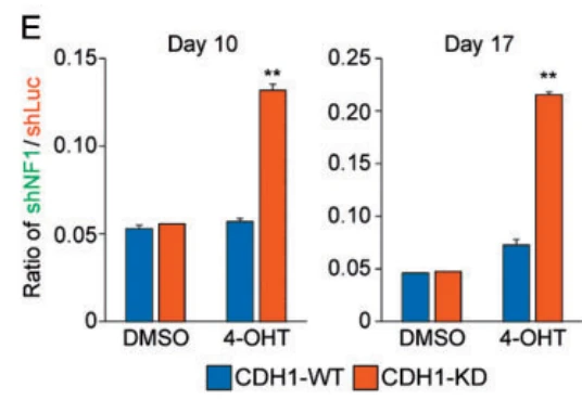
In the CDH1-WT background, NF1 knockdown did not confer a significant competitive growth advantage over parental cells under 4-OHT treatment across 10 or 17 days, with the ratio of shNF1/shLuc cells remaining low.
Effect of NF1 knockdown on competitive growth under 4-OHT treatment in CDH1-KD background
Assay
Competitive co-culture assay (flow cytometry)
Pre-treatment Protocol
T47D cells transduced to knock down CDH1 (shECad) or control (shCntrl), followed by NF1 knockdown labeled with GFP (shNF1-GFP) or control labeled with mCherry (shLuc-mCherry); detailed shRNA sequences and delivery reagents not described
Treatment
4-OHT
Incubation Time
10 days, 17 days
Control Setup
Solvent control (DMSO), CDH1-WT co-culture control
Results
In CDH1-KD cells, NF1 knockdown conferred a competitive advantage upon 4-OHT treatment, increasing from 5% of the population to 13% at 10 days and 21% at 17 days (p < 0.00001), whereas this advantage was absent in solvent control and CDH1-WT cells.
Floxed beta-actin mice crossed to Cagg-CreER · Immunofluorescence
Research Field
Cell Biology
Sample
Floxed beta-actin mice crossed to Cagg-CreER
Research Purpose
Treadmilling assessment via conditional beta-actin knockout
Assay
Immunofluorescence
Treatment
Tamoxifen
Control Setup
Floxed beta-actin mice crossed to Cagg-CreER without tamoxifen
Results
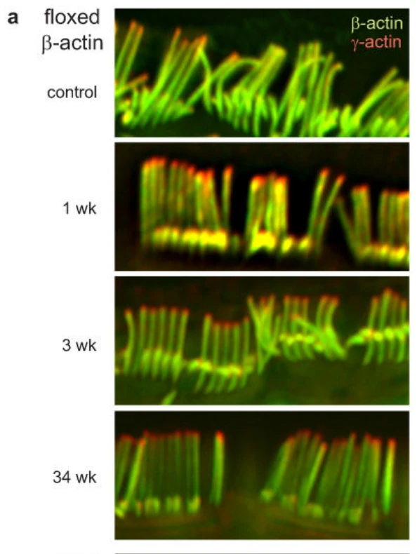
Following tamoxifen-induced β-actin excision in 3-week-old mice, loss of β-actin protein was confined to a ~0.4-μm region at the stereocilia tips at 1, 3, and 34 weeks, while the remaining stereocilia shaft retained β-actin.
Floxed gamma-actin mice crossed to Cagg-CreER (cochlear inner hair cells)
Research Purpose
Treadmilling assessment via conditional gamma-actin knockout
Assay
Immunofluorescence
Treatment
Tamoxifen
Incubation Time
18 weeks
Control Setup
Untreated floxed mice without tamoxifen
Results
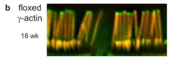
In mice carrying floxed Actg1 crossed with CreER, tamoxifen-induced gene deletion resulted in loss of gamma-actin only at the distal ~0.4 µm tips of stereocilia after 18 weeks, with gamma-actin retained throughout the shaft without progressive loss toward the base.
Primary T-lymphoma cells · Cell Titer-Blue Viability Assay, Western Blot
Research Field
Oncology
Sample
Primary T-lymphoma cells
Research Purpose
In vitro mutp53 ablation and cell death induction
Assay
Cell Titer-Blue Viability Assay, Western Blot
Culture Conditions
Lymphoma medium (1:1 DMEM/IMDM supplemented with 10% FBS, L-glutamine, Penicillin/Streptomycin, Antibiotic/Antimycotic, β-mercaptoethanol)
Seeding Density
15×10^6 live cells per 6 cm dish
Working Concentration
1 µM
Treatment
4-hydroxytamoxifen
Incubation Time
3–6 days
Control Setup
Untreated, vehicle (EtOH), and Q/-;ERT2/+ control cells
Results
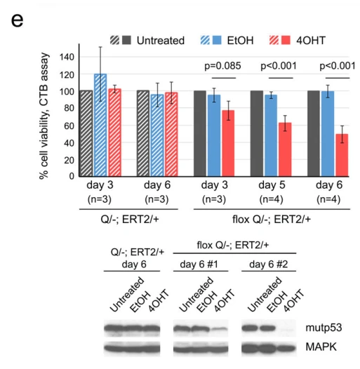
Treatment of floxQ/-;ERT2/+ T-lymphoma cells with 1 µM 4OHT caused significant loss of cell viability relative to untreated or EtOH controls at day 3 (p=0.065), day 5 (p<0.001), and day 6 (p<0.001) alongside mutp53 protein depletion, whereas Q/-;ERT2/+ control cells maintained viability and mutp53 levels.
Primary T-lymphoma cells · Cell Titer-Blue Viability Assay, Western Blot
Research Field
Oncology
Sample
Primary T-lymphoma cells
Research Purpose
In vitro mutp53 ablation control
Assay
Cell Titer-Blue Viability Assay, Western Blot
Culture Conditions
Lymphoma medium (1:1 DMEM/IMDM supplemented with 10% FBS, L-glutamine, Penicillin/Streptomycin, Antibiotic/Antimycotic, β-mercaptoethanol)
Seeding Density
15×10^6 live cells per 6 cm dish
Working Concentration
1 µM
Treatment
4OHT
Incubation Time
3–6 days
Control Setup
Untreated, vehicle (EtOH)
Results
In control Q/-;ERT2/+ primary T-lymphoma cells lacking the floxed allele, 4OHT treatment did not induce cell death or alter cell viability at day 3 or day 6 (p=0.085 at day 6), and sustained high mutp53 expression was maintained as shown by Western blot.
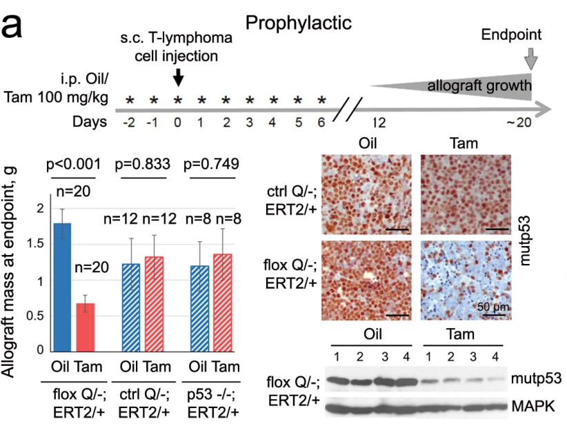
Prophylactic in vivo mutp53 ablation in s.c. allografts
Assay
Tumor weight, immunohistochemistry, and immunoblotting
Working Concentration
100 mg/kg
Treatment
Tamoxifen
Incubation Time
9 days
Control Setup
Corn oil vehicle; ctrl Q/-;ERT2/+ and p53-/-;ERT2/+ allografts treated with Tamoxifen or oil
Results
Prophylactic Tamoxifen treatment significantly curbed tumor growth and reduced endpoint allograft mass specifically in floxQ/-;ERT2/+ tumors (p<0.001, n=20), but had no inhibitory effect on ctrl Q/-;ERT2/+ (p=0.833) or p53-/-;ERT2/+ (p=0.749) control allografts.
Tamoxifen 100 mg/kg i.p. daily for 2 days prior to lymphoma cell transplantation (or corn oil vehicle); s.c. inoculation with 10^5 non-floxed control Q/-; Rosa26CreERT2/+ primary T-lymphoma cells per site suspended in 3:1 PBS/Matrigel at 4 dorsal sites on day 0
Treatment
Tamoxifen
Incubation Time
7 days
Control Setup
corn oil vehicle (Oil), n=12 allografts
Results
In non-inactivatable control Q/-;ERT2/+ allografts, prophylactic Tamoxifen treatment produced no significant reduction in tumor weight at endpoint compared to vehicle (p=0.833, n=8 for Tam vs n=12 for Oil).
Prophylactic in vivo mutp53 ablation control in s.c. allografts
Assay
Tumor Weight
Working Concentration
100 mg/kg
Pre-treatment Protocol
Subcutaneous transplantation of 10^5 primary p53-/-;ERT2/+ T-lymphoma cells per site suspended in 3:1 PBS/Matrigel into four dorsal sites of 6-7 weeks old male Nu/Nu mice on day 0 (two days after start of Tamoxifen treatment)
Treatment
Tamoxifen
Incubation Time
9 days (daily i.p. injections from day -2 to day 6)
Control Setup
Corn oil vehicle control (n=8 allografts)
Results
Prophylactic administration of Tamoxifen did not alter endpoint allograft tumor mass in p53-/-;ERT2/+ allograft controls compared to corn oil vehicle (p=0.749, n=8 allografts per group).
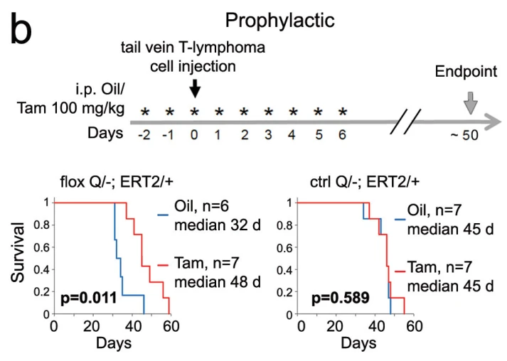
Nude mice injected intravenously with floxQ/-;ERT2/+ T-lymphoma cells
Research Purpose
Prophylactic in vivo mutp53 ablation in intravenous allografts
Assay
Kaplan-Meier Survival Analysis
Working Concentration
100 mg/kg
Pre-treatment Protocol
Tamoxifen 100 mg/kg i.p. daily for 2 days prior to intravenous lymphoma cell transplantation
Treatment
Tamoxifen
Incubation Time
9 days (daily i.p. injections across day -2 to day 6)
Control Setup
Corn oil vehicle control; non-floxed Q/-;ERT2/+ control allografts treated with oil or Tamoxifen
Results
Prophylactic ablation of mutp53 significantly extended median survival from 32 days in oil-treated controls (n=6) to 48 days in Tamoxifen-treated recipients (n=7, p=0.011), whereas Tamoxifen exerted no survival benefit in recipient mice bearing control non-inactivatable Q/-;ERT2/+ lymphoma cells (median 45 days in both oil and Tam groups, p=0.589).
Nude mice injected intravenously with ctrl Q/-;ERT2/+ T-lymphoma cells
Research Purpose
Prophylactic in vivo mutp53 ablation control in intravenous allografts
Assay
Kaplan-Meier survival analysis
Working Concentration
100 mg/kg
Treatment
Tamoxifen
Incubation Time
9 daily doses
Control Setup
Corn oil vehicle control
Results
Tamoxifen treatment showed no significant difference in survival compared to oil in recipients transplanted with ctrl Q/-;ERT2/+ T-lymphoma cells (median 45 days in both groups, p=0.589).
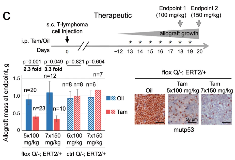
Therapeutic in vivo mutp53 ablation in s.c. allografts
Assay
Tumor Mass Measurement, Caliper Tumor Volume Measurement, Immunohistochemistry
Seeding Density
10^5 cells/site at 4 dorsal sites in 3:1 PBS/Matrigel
Working Concentration
100 mg/kg, 150 mg/kg
Treatment
Tamoxifen
Incubation Time
5 to 7 daily injections
Control Setup
corn oil vehicle, constitutive Q/-;ERT2/+ control allografts, p53-/-;ERT2/+ control allografts
Results
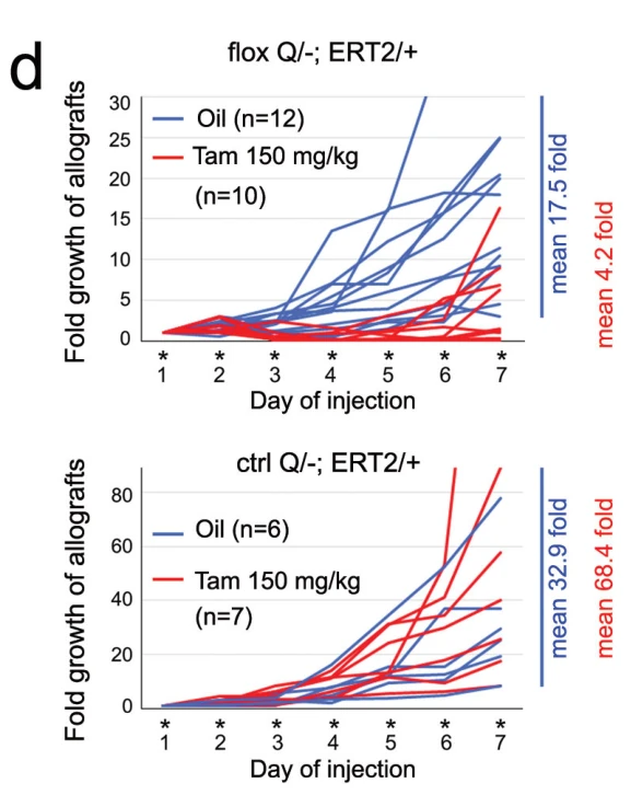
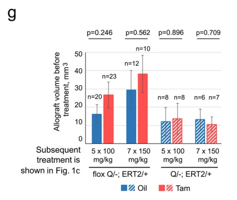
Therapeutic Tamoxifen administration curbed allograft growth in a dose-dependent manner (mean fold growth 4.2 fold with 150 mg/kg Tamoxifen vs 17.5 fold with oil; endpoint tumor mass reduced from 0.77 g to 0.33 g, 2.3-fold reduction with 5x100 mg/kg, p=0.001; and reduced from 1.05 g to 0.32 g, 3.3-fold reduction with 7x150 mg/kg, p=0.049), with no response observed in p53-null controls.
Therapeutic in vivo mutp53 ablation control in s.c. allografts
Assay
Tumor mass, fold growth, and allograft volume
Seeding Density
10^5 lymphoma cells per site in 3:1 PBS/Matrigel (4 dorsal sites per mouse)
Working Concentration
100 mg/kg, 150 mg/kg
Treatment
Tamoxifen
Incubation Time
5 to 7 days
Control Setup
Corn oil vehicle
Results
In control Q/-;ERT2/+ allografts lacking the floxed allele, therapeutic Tamoxifen administration did not significantly reduce tumor mass at endpoint under either 100 mg/kg for 5 days (p=0.821) or 150 mg/kg for 7 days (p=0.604). Allografts displayed strong fold growth reaching a mean of 68.4-fold after 7 days of 150 mg/kg Tamoxifen treatment, closely tracking oil-treated controls (mean 32.9-fold).
Therapeutic in vivo mutp53 ablation control in s.c. allografts
Assay
Caliper Measurement, Tumor Weight Measurement
Seeding Density
10^5 cells per site in 3:1 PBS/Matrigel (4 dorsal sites per mouse)
Working Concentration
150 mg/kg
Treatment
Tamoxifen
Incubation Time
7 days
Control Setup
corn oil vehicle
Results
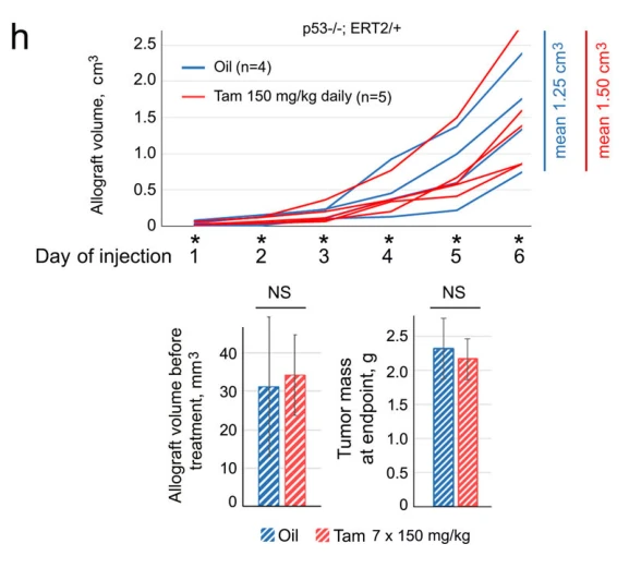
Tamoxifen treatment showed no significant inhibitory effect on p53-/-;ERT2/+ allograft volume over time or tumor mass at endpoint (1.50 cm3 in Tam vs 1.25 cm3 in Oil, NS), confirming the specificity of therapeutic ablation.
Therapeutic mutp53 ablation in SCID mice bearing s.c. allografts
Assay
Survival Analysis, Immunofluorescence
Working Concentration
100 mg/kg
Pre-treatment Protocol
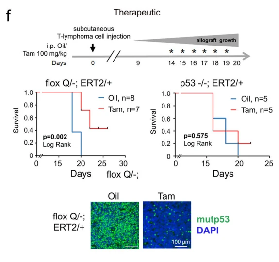
Primary T-lymphoma cells isolated from floxQ/-;ERT2/+ or Q/-;ERT2/+ donor mice were subcutaneously transplanted into SCID recipients, and tumors were allowed to become visible before starting treatment
Treatment
Tamoxifen
Incubation Time
daily until endpoint (5 to 7 daily injections)
Control Setup
Corn oil vehicle control, non-switchable Q/-;ERT2/+ control allografts
FloxQ/-;ERT2/+ mice with clinically advanced autochthonous tumors · Ultrasound, Immunohistochemistry, and H&E Staining
Research Field
Oncology
Sample
FloxQ/-;ERT2/+ mice with clinically advanced autochthonous tumors
Research Purpose
Therapeutic mutp53 ablation in clinically advanced autochthonous tumors
Assay
Ultrasound, Immunohistochemistry, and H&E Staining
Working Concentration
150 mg/kg daily for 7 days, followed by 100 mg/kg three times a week
Treatment
Tamoxifen
Incubation Time
until sacrifice (endpoint)
Control Setup
corn oil vehicle, Q/-;ERT2/+ control mice treated with Tamoxifen
Results
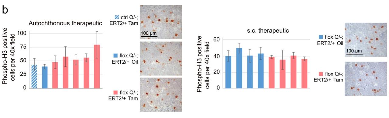
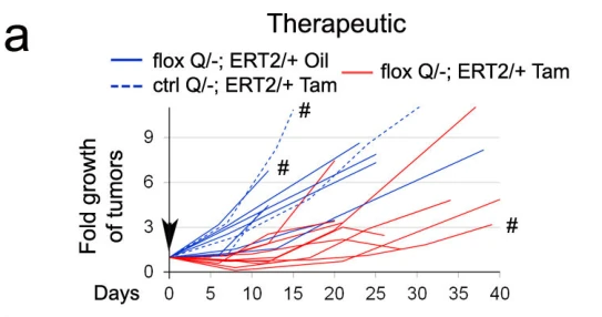
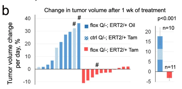
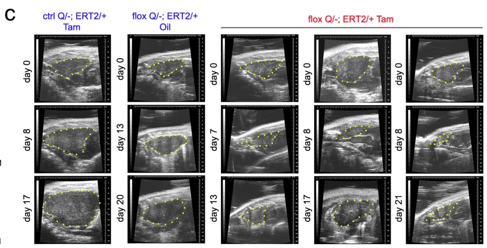
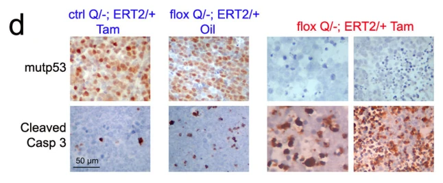
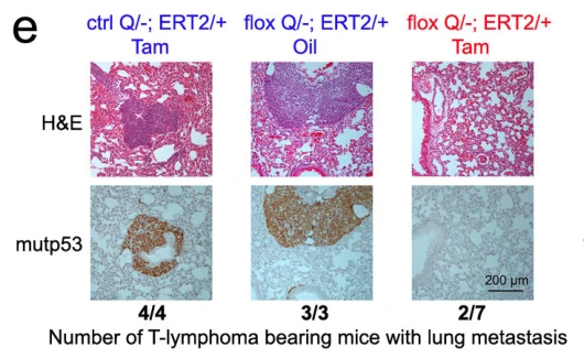
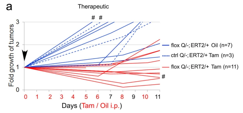
Clinically advanced autochthonous tumors in floxQ/-;ERT2/+ mice responded to Tamoxifen-induced mutp53 deletion with marked regression or stagnation (p<0.001 vs controls), driven by pronounced tumor apoptosis (cleaved caspase 3 induction) without significant change in mitotic index, and accompanied by strong suppression of lung metastasis (metastases in 2/7 Tam-treated vs 3/3 oil-treated floxQ/-;ERT2/+ mice).
Therapeutic mutp53 ablation control in clinically advanced autochthonous tumors
Assay
Ultrasound, Daily Tumor Growth Rate Calculation, Immunohistochemistry, and Metastasis Evaluation
Working Concentration
150 mg/kg daily for 7 days, then 100 mg/kg three times weekly
Treatment
Tamoxifen
Incubation Time
until sacrifice
Control Setup
floxQ/-;ERT2/+ mice treated with corn oil, floxQ/-;ERT2/+ mice treated with Tamoxifen
Results
In non-switchable control Q/-;ERT2/+ mice bearing clinically advanced tumors, tamoxifen treatment failed to induce tumor regression or stagnation, with tumors exhibiting rapid positive daily growth (~15-20% per day) similar to oil-treated floxQ/- controls (p<0.001 vs floxQ/- Tam), retained strong mutp53 expression without significant apoptosis, and developed widespread lung metastases (4/4 mice positive).
Mutp53 ablation in early organ-confined autochthonous disease
Assay
Survival Analysis, Immunohistochemistry
Working Concentration
100 mg/kg
Treatment
Tamoxifen
Incubation Time
5 days
Control Setup
Corn oil vehicle control
Results
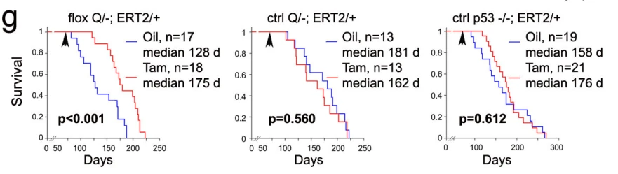
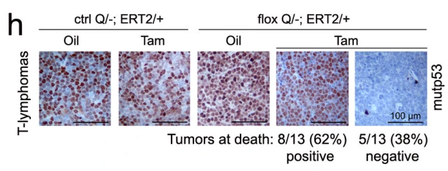
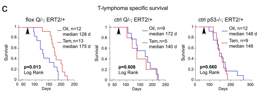
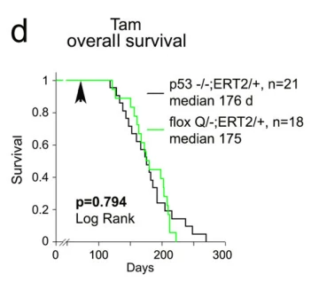
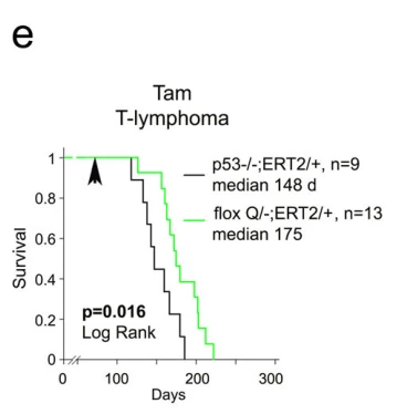
Tamoxifen-induced mutp53 ablation at 10 weeks of age extended median overall and T-lymphoma-specific survival by 37% from 128 to 175 days (p<0.001); at endpoint, 74% (17/23) of recurrent tumors re-expressed stabilized mutp53.
Mutp53 ablation control in early autochthonous disease
Assay
Kaplan-Meier survival analysis
Working Concentration
100 mg/kg
Treatment
Tamoxifen
Incubation Time
5 consecutive days
Control Setup
Oil vehicle (corn oil alone)
Results
In control p53-/-;ERT2/+ mice treated at 10 weeks of age, Tamoxifen did not significantly alter cancer-related overall survival (median 176 days vs 158 days for oil, p=0.612, Fig. 2g; median 148 days for both Tam and oil, p=0.660, Extended Data Fig. 2c) or T-lymphoma-specific survival, confirming that the therapeutic survival benefit of Tamoxifen-induced Cre recombination is strictly mutp53-dependent.
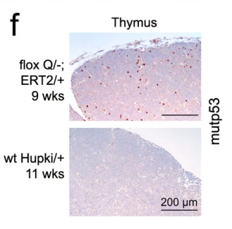
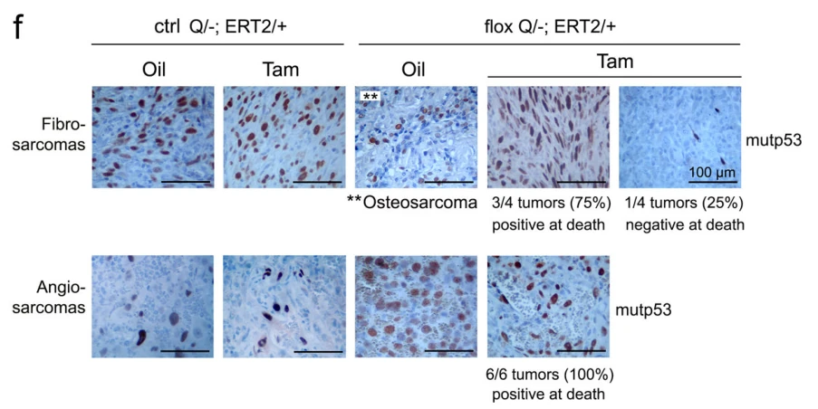
In non-inactivatable constitutive ctrl Q/-;ERT2/+ control mice treated at 10 weeks of age, Tamoxifen did not alter overall survival compared with oil (median 162 d vs 181 d, p=0.560) or T-lymphoma-specific survival (median 140 d vs 172 d, p=0.608), and endpoint sarcomas maintained positive mutp53 immunostaining.