
-
 Australia
-
 Austria
-
 Belgium
-
 Brazil
-
 Canada
-
 China
China
-
 Czech Republic
-
 Denmark
-
 Finland
-
 France
-
 Germany
-
 Greece
-
 Hong Kong
-
 Hungary
-
 Iceland
-
 India
India
-
 Ireland
-
 Israel
-
 Italy
Italy
-
 Japan
Japan
-
 Korea
Korea
-
 Luxembourg
-
 Malaysia
-
 Netherlands
-
 New Zealand
-
 Norway
-
 Poland
-
 Qatar
Qatar
-
 Romania
-
 Saudi Arabia
-
 Singapore
-
 Spain
Spain
-
 Sweden
-
 Switzerland
-
 Taiwan
-
 Turkey
-
 United Kingdom
-
 United States
research use only
TMEM119 (Microglial Marker) Antibody (Rabbit mAb) [H12P23]
CatNo: F1546
-
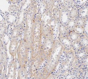 Immunohistochemical analysis of formalin fixed paraffin embedded mouse kidney tissue with F1546 at 1:200 dilution.
Usage Information
| Dilution |
|---|
|
| Application |
|---|
| IHC |
| Reactivity |
|---|
| Mouse |
| Source |
|---|
| Rabbit Monoclonal Antibody |
| Storage Buffer |
|---|
| PBS, pH 7.2+50% Glycerol+0.05% BSA+0.01% NaN3 |
| Storage (from the date of receipt) |
|---|
| -20°C (avoid freeze-thaw cycles), 2 years |
| Predicted MW |
|---|
| 29 kDa |
| Positive Control | Mouse cerebrum tissue; Mouse corpus callosum tissue; Normal mouse brain tissue |
|---|---|
| Negative Control |
Experimental Methods
| IHC |
|---|
Experimental Protocol:
Deparaffinization/Rehydration
1. Deparaffinize/hydrate sections:
2. Incubate sections in three washes of xylene for 5 min each.
3. Incubate sections in two washes of 100% ethanol for 10 min each.
4. Incubate sections in two washes of 95% ethanol for 10 min each.
5. Wash sections two times in dH2O for 5 min each.
6.Antigen retrieval: For Citrate: Heat slides in a microwave submersed in 1X citrate unmasking solution until boiling is initiated; continue with 10 min at a sub-boiling temperature (95°-98°C). Cool slides on bench top for 30 min.
Staining
1. Wash sections in dH2O three times for 5 min each.
2. Incubate sections in 3% hydrogen peroxide for 10 min.
3. Wash sections in dH2O two times for 5 min each.
4. Wash sections in wash buffer for 5 min.
5. Block each section with 100–400 µl of blocking solution for 1 hr at room temperature.
6. Remove blocking solution and add 100–400 µl primary antibody diluent in to each section. Incubate overnight at 4°C.
7. Remove antibody solution and wash sections with wash buffer three times for 5 min each.
8. Cover section with 1–3 drops HRPas needed. Incubate in a humidified chamber for 30 min at room temperature.
9. Wash sections three times with wash buffer for 5 min each.
10. Add DAB Chromogen Concentrate to DAB Diluent and mix well before use.
11. Apply 100–400 µl DAB to each section and monitor closely. 1–10 min generally provides an acceptable staining intensity.
12. Immerse slides in dH2O.
13. If desired, counterstain sections with hematoxylin.
14. Wash sections in dH2O two times for 5 min each.
15. Dehydrate sections: Incubate sections in 95% ethanol two times for 10 sec each; Repeat in 100% ethanol, incubating sections two times for 10 sec each; Repeat in xylene, incubating sections two times for 10 sec each.
16. Mount sections with coverslips and mounting medium.
|
Biological Description
| Specificity |
|---|
TMEM119 (Microglial Marker) Antibody (Rabbit mAb) [H12P23] detects endogenous levels of total TMEM119 (Microglial Marker) protein. |
| Uniprot ID |
|---|
| Q4V9L6 |
| Clone |
|---|
| H12P23 |
| Synonym(s) |
|---|
| Transmembrane protein 119, Osteoblast induction factor, OBIF, Tmem119 |
| Background |
|---|
TMEM119 is a type I transmembrane protein of unknown enzymatic activity that is highly enriched in parenchymal microglia and has emerged as a robust marker for resident microglia in the central nervous system, where it helps distinguish yolk sac–derived microglia from infiltrating blood‑borne macrophages in many physiological and pathological contexts. The protein contains a luminal/extracellular N‑terminal region, a single transmembrane segment, and a short cytoplasmic tail, and its expression is largely restricted to microglia within brain parenchyma, with minimal or absent expression in neurons, astrocytes, oligodendrocytes, and most peripheral myeloid cells, which allows its use as a relatively specific microglial surface marker in immunohistochemistry, flow cytometry, and transcriptomic targeting approaches. Comparative microglial transcriptome analyses identified TMEM119 as a conserved microglial signature gene whose mRNA and protein are maintained across resting and classically activated states more stably than some other markers, and TMEM119 immunoreactivity labels IBA1⁺CD68⁺ microglia in human neurodegenerative brains while sparsely labeling or failing to label IBA1⁺ macrophages infiltrating multiple sclerosis lesions or infarcts, which supports its use to discriminate resident microglia from recruited monocyte-derived macrophages in human tissue. TMEM119 expression changes with microglial activation state and anatomical location: traumatic brain injury, experimental demyelination, ischemia, and other acute or chronic insults are often accompanied by decreased TMEM119 signal in microglia that adopt amoeboid or DAM‑like phenotypes, and TMEM119 protein is reduced particularly in lesion cores and periplaque regions while being better preserved in more distal or less activated microglial populations, indicating that TMEM119 marks homeostatic microglia and becomes downregulated as microglia transition to strongly reactive states. Recent work shows that TMEM119 can bind amyloid‑β oligomers, participate in recruitment of low-density lipoprotein receptor family members such as LRP1, and influence amyloid clearance and microglial phagocytic capacity in Alzheimer’s disease models, suggesting a functional role in microglial handling of aggregated proteins in addition to its value as a marker. TMEM119 mRNA levels are increased in some Alzheimer’s disease brains while protein levels show region- and context-dependent changes, and TMEM119-positive microglia do not consistently align with classical M1 or M2 polarization markers, which positions TMEM119 as a marker of microglial lineage and homeostatic identity rather than of a simple activation state. Expression outside the CNS has been reported in select peripheral cell types under specific conditions, and TMEM119 can be downregulated in highly stressed or disease-associated microglia, so its specificity and sensitivity depend on disease model and time point; these features are critical when TMEM119 promoter-driven reporter lines or antibody-based quantification are used to define or isolate microglial populations. |
| References |
|---|
|
Tech Support
Tel: +1-832-582-8158 Ext:3
If you have any other enquiries, please leave a message.

Products are for research use only. Not for human use. We do not sell to patients.
©Copyright 2013 Selleck Chemicals. All Rights Reserved.