
-
 Australia
-
 Austria
-
 Belgium
-
 Brazil
-
 Canada
-
 China
China
-
 Czech Republic
-
 Denmark
-
 Finland
-
 France
-
 Germany
-
 Greece
-
 Hong Kong
-
 Hungary
-
 Iceland
-
 India
India
-
 Ireland
-
 Israel
-
 Italy
Italy
-
 Japan
Japan
-
 Korea
Korea
-
 Luxembourg
-
 Malaysia
-
 Netherlands
-
 New Zealand
-
 Norway
-
 Poland
-
 Qatar
Qatar
-
 Romania
-
 Saudi Arabia
-
 Singapore
-
 Spain
Spain
-
 Sweden
-
 Switzerland
-
 Taiwan
-
 Turkey
-
 United Kingdom
-
 United States
research use only
TER-119 Antibody (Rat mAb) [H18E16]
CatNo: F5781
-
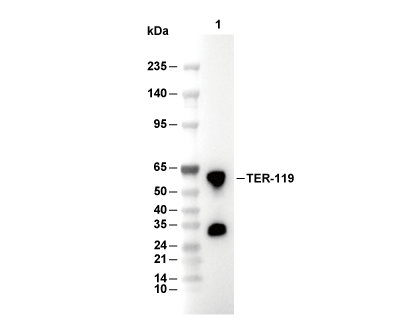 Lane 1: Mouse spleen
Experiment Essentials
This antibody requires an anti-rat secondary antibody.
Usage Information
| Dilution |
|---|
|
| Application |
|---|
| WB, FCM |
| Reactivity |
|---|
| Mouse |
| Source |
|---|
| Rat Monoclonal Antibody |
| Storage Buffer |
|---|
| PBS, pH 7.2+50% Glycerol+0.05% BSA+0.01% NaN3 |
| Storage (from the date of receipt) |
|---|
| -20°C (avoid freeze-thaw cycles), 2 years |
| Positive Control | C57BL/6 bone marrow cells |
|---|---|
| Negative Control |
Experimental Methods
| WB |
|---|
Experimental Protocol:
Sample preparation
1. Tissue: Lyse the tissue sample by adding an appropriate volume of ice-cold Lysis Buffer (containing Protease Inhibitor Cocktail),and homogenize the tissue at a low temperature.
2. Adherent cell: Aspirate the culture medium and wash the cells with ice-cold PBS twice. Lyse the cells by adding an appropriate volume of Lysis Buffer (containing Protease Inhibitor Cocktail) and put the sample on ice for 5 min.
3. Suspension cell: Transfer the culture medium to a pre-cooled centrifuge tube. Centrifuge and aspirate the supernatant. Wash the cells with ice-cold PBS twice. Lyse the cells by adding an appropriate volume of Lysis Buffer (containing Protease Inhibitor Cocktail) and put the sample on ice for 5 min.
4. Place the lysate into a pre-cooled microcentrifuge tube. Centrifuge at 4°C for 15 min. Collect the supernatant;
5. Remove a small volume of lysate to determine the protein concentration;
6. Combine the lysate with protein loading buffer. Boil 20 µL sample under 95-100°C for 5 min. Centrifuge for 5 min after cool down on ice.
Electrophoretic separation
1. According to the concentration of extracted protein, load appropriate amount of protein sample and marker onto SDS-PAGE gels for electrophoresis. Reference Table for Selecting SDS-PAGE Separation Gel Concentrations
2. Power up 80V for 30 minutes. Then the power supply is adjusted (110 V~150 V), the Marker is observed, and the electrophoresis can be stopped when the indicator band of the predyed protein Marker where the protein is located is properly separated. (Note that the current should not be too large when electrophoresis, too large current (more than 150 mA) will cause the temperature to rise, affecting the result of running glue. If high currents cannot be avoided, an ice bath can be used to cool the bath.)
Transfer membrane
1. Take out the converter, soak the clip and consumables in the pre-cooled converter;
2. Activate PVDF membrane with methanol for 1 min and rinse with transfer buffer;
3. Install it in the order of "black edge of clip - sponge - filter paper - filter paper - glue -PVDF membrane - filter paper - filter paper - sponge - white edge of clip";
4. The protein was electrotransferred to PVDF membrane. Reference Table for Selecting PVDF Membrane Pore Size Specifications
( Note that the transfer conditions can be adjusted according to the protein size. For high-molecular-weight proteins, a higher current and longer transfer time are recommended. However, ensure that the transfer tank remains at a low temperature to prevent gel melting.)
Block
1. After electrotransfer, wash the film with TBST at room temperature for 5 minutes;
2. Incubate the film in the blocking solution for 1 hour at room temperature;
3. Wash the film with TBST for 3 times, 5 minutes each time.
Antibody incubation
1. Use 5% skim milk powder to prepare the primary antibody working liquid (recommended dilution ratio for primary antibody 1:1000), gently shake and incubate with the film at 4°C overnight;
2. Wash the film with TBST 3 times, 5 minutes each time;
3. Add the secondary antibody to the blocking solution and incubate with the film gently at room temperature for 1 hour;
4. After incubation, wash the film with TBST 3 times for 5 minutes each time.
Antibody staining
1. Add the prepared ECL luminescent substrate (or select other color developing substrate according to the second antibody) and mix evenly;
2. Incubate with the film for 1 minute, remove excess substrate (keep the film moist), wrap with plastic film, and expose in the imaging system.
|
Biological Description
| Specificity |
|---|
TER-119 Antibody (Rat mAb) [H18E16] detects mouse erythroid-specific 52 kDa glycophorin A-associated antigen. |
| Clone |
|---|
| H18E16 |
| Synonym(s) |
|---|
| Ly-76; Ly76; TER119 |
| Background |
|---|
TER‑119 is a rat monoclonal antibody that recognizes a 52 kDa erythroid‑specific cell surface antigen associated with glycophorin A, and it serves as a robust lineage marker for murine erythroid cells from the proerythroblast stage through mature erythrocytes in embryonic yolk sac, fetal and newborn liver, adult bone marrow, peripheral blood, and spleen. The TER‑119 antigen is not detectable on earlier erythroid progenitors with BFU‑E or CFU‑E activity, nor on hematopoietic stem cells, lymphoid or myeloid cells, or erythroleukemia lines, which allows selective identification and depletion of committed erythroid cells in hematopoietic assays and flow cytometric “lineage cocktail” panels. Biochemical and molecular analyses show that the epitope recognized by TER‑119 is a glycophorin A–associated glycoprotein, sometimes referred to as Ly‑76, that forms part of a membrane protein complex linked to band 3 and other cytoskeletal components, implicating this antigen cluster in maintenance of erythrocyte membrane integrity, mechanical flexibility, and proper ion transport in circulating red cells. Functionally, TER‑119 staining resolves successive stages of erythroid maturation, enabling quantitative assessment of erythropoiesis, detection of defects in red cell development, and monitoring of responses to erythropoietic stimuli or suppressive interventions in mouse models of anemia, hemolysis, and bone marrow failure. Because TER‑119 recognizes a conformational glyco‑epitope dependent on the organization of glycophorin A–associated membrane proteins and sialylated structures, it is widely used in flow cytometry, immunohistochemistry, and immunofluorescence to label and enumerate erythroid lineage cells, but the antigen itself is not typically considered a signaling receptor or direct therapeutic target. |
| References |
|---|
|
Tech Support
Tel: +1-832-582-8158 Ext:3
If you have any other enquiries, please leave a message.

Products are for research use only. Not for human use. We do not sell to patients.
©Copyright 2013 Selleck Chemicals. All Rights Reserved.