-
 Australia
-
 Austria
-
 Belgium
-
 Brazil
-
 Canada
-
 China
China
-
 Czech Republic
-
 Denmark
-
 Finland
-
 France
-
 Germany
-
 Greece
-
 Hong Kong
-
 Hungary
-
 Iceland
-
 India
India
-
 Ireland
-
 Israel
-
 Italy
Italy
-
 Japan
Japan
-
 Korea
Korea
-
 Luxembourg
-
 Malaysia
-
 Netherlands
-
 New Zealand
-
 Norway
-
 Poland
-
 Qatar
Qatar
-
 Romania
-
 Saudi Arabia
-
 Singapore
-
 Spain
Spain
-
 Sweden
-
 Switzerland
-
 Taiwan
-
 Turkey
-
 United Kingdom
-
 United States
research use only
TBC1D7 Antibody [D5H17]
Cat.No.: F5142
Application:
Reactivity:
-
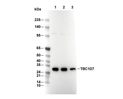 Lane 1: 3T3, Lane 2: Hela, Lane 3: C2C12
Experiment Essentials
WB
Recommended wet transfer conditions: 200 mA, 60 min.
Recommended wet transfer conditions: 200 mA, 60 min.
Usage Information
| Dilution |
|---|
|
| Application |
|---|
| WB, IP |
| Reactivity |
|---|
| Human, Mouse, Rat |
| Source |
|---|
| Rabbit Monoclonal Antibody |
| Storage Buffer |
|---|
| PBS, pH 7.2+50% Glycerol+0.05% BSA+0.01% NaN3 |
| Storage (from the date of receipt) |
|---|
| -20°C (avoid freeze-thaw cycles), 2 years |
| Predicted MW |
|---|
| 30 kDa |
| Positive Control | NIH/3T3 cells; HeLa cells; C2C12 cells |
|---|---|
| Negative Control |
Experimental Methods
| WB |
|---|
Experimental Protocol:
Sample preparation
1. Tissue: Lyse the tissue sample by adding an appropriate volume of ice-cold RIPA/NP-40 Lysis Buffer (containing Protease Inhibitor Cocktail),and homogenize the tissue at a low temperature. 2. Adherent cell: Aspirate the culture medium and wash the cells with ice-cold PBS twice. Lyse the cells by adding an appropriate volume of RIPA/NP-40 Lysis Buffer (containing Protease Inhibitor Cocktail) and put the sample on ice for 5 min. 3. Suspension cell: Transfer the culture medium to a pre-cooled centrifuge tube. Centrifuge and aspirate the supernatant. Wash the cells with ice-cold PBS twice. Lyse the cells by adding an appropriate volume of RIPA/NP-40 Lysis Buffer (containing Protease Inhibitor Cocktail) and put the sample on ice for 5 min. 4. Place the lysate into a pre-cooled microcentrifuge tube. Centrifuge at 4°C for 15 min. Collect the supernatant;
5. Remove a small volume of lysate to determine the protein concentration;
6. Combine the lysate with protein loading buffer. Boil 20 µL sample under 95-100°C for 5 min. Centrifuge for 5 min after cool down on ice.
Electrophoretic separation
1. According to the concentration of extracted protein, load appropriate amount of protein sample and marker onto SDS-PAGE gels for electrophoresis. Recommended separating gel (lower gel) concentration: 10%. Reference Table for Selecting SDS-PAGE Separation Gel Concentrations 2. Power up 80V for 30 minutes. Then the power supply is adjusted (110 V~150 V), the Marker is observed, and the electrophoresis can be stopped when the indicator band of the predyed protein Marker where the protein is located is properly separated. (Note that the current should not be too large when electrophoresis, too large current (more than 150 mA) will cause the temperature to rise, affecting the result of running glue. If high currents cannot be avoided, an ice bath can be used to cool the bath.)
Transfer membrane
1. Take out the converter, soak the clip and consumables in the pre-cooled converter;
2. Activate PVDF membrane with methanol for 1 min and rinse with transfer buffer;
3. Install it in the order of "black edge of clip - sponge - filter paper - filter paper - glue -PVDF membrane - filter paper - filter paper - sponge - white edge of clip"; 4. The protein was electrotransferred to PVDF membrane. ( 0.45 µm PVDF membrane is recommended ) Reference Table for Selecting PVDF Membrane Pore Size Specifications Recommended conditions for wet transfer: 200 mA, 60 min. ( Note that the transfer conditions can be adjusted according to the protein size. For high-molecular-weight proteins, a higher current and longer transfer time are recommended. However, ensure that the transfer tank remains at a low temperature to prevent gel melting.)
Block
1. After electrotransfer, wash the film with TBST at room temperature for 5 minutes;
2. Incubate the film in the blocking solution for 1 hour at room temperature;
3. Wash the film with TBST for 3 times, 5 minutes each time.
Antibody incubation
1. Use 5% skim milk powder to prepare the primary antibody working liquid (recommended dilution ratio for primary antibody 1:1000), gently shake and incubate with the film at 4°C overnight; 2. Wash the film with TBST 3 times, 5 minutes each time;
3. Add the secondary antibody to the blocking solution and incubate with the film gently at room temperature for 1 hour;
4. After incubation, wash the film with TBST 3 times for 5 minutes each time.
Antibody staining
1. Add the prepared ECL luminescent substrate (or select other color developing substrate according to the second antibody) and mix evenly;
2. Incubate with the film for 1 minute, remove excess substrate (keep the film moist), wrap with plastic film, and expose in the imaging system. |
Biological Description
| Specificity |
|---|
| TBC1D7 Antibody [D5H17] detects endogenous levels of total TBC1D7 protein. |
| Subcellular Location |
|---|
| Cytoplasm, Cytoplasmic vesicle, Lysosome, Membrane |
| Uniprot ID |
|---|
| Q9P0N9 |
| Clone |
|---|
| D5H17 |
| Synonym(s) |
|---|
| TBC1 domain family member 7; Cell migration-inducing protein 23; TBC1D7; TBC7; HSPC239 |
| Background |
|---|
| TBC1D7, a non-canonical member of the TBC/RabGAP protein family that lacks the typical catalytic arginine and glutamine fingers, serves as the third essential subunit of the TSC1-TSC2 complex, known as the TSC complex. By binding to the C-terminal coiled-coil domain of TSC1, TBC1D7 stabilizes the heterodimerization of TSC1 and TSC2 and enhances the Rheb-GAP activity required for effective mTORC1 suppression. TBC1D7 adopts a compact, globular fold composed of fifteen α-helices, with conserved residues on α4 and α5 mediating interaction with TSC1 through a 2:2 heterotetramer interface, where the C-terminal tip of α4 crosslinks adjacent TSC1 molecules, reinforcing the rigidity and stability of the complex. Phosphorylation of Ser124 by Akt enables recruitment of 14-3-3 proteins, further stabilizing TBC1D7. TBC1D7 is crucial for maintaining the integrity of the TSC complex, preventing TSC1 dissociation, and amplifying TSC2-mediated Rheb-GTP hydrolysis, thereby dampening mTORC1-driven anabolic pathways such as protein synthesis and cell growth and limiting lysosome-based mTORC1 activation during nutrient stress. TBC1D7 also fine-tunes autophagy, insulin and PI3K signaling responses, and systemic growth, with some functions occurring independently of the TSC complex. Loss of TBC1D7 destabilizes the TSC1-TSC2 complex, increases Rheb-GTP and mTORC1 activity, leads to cell enlargement, and delays autophagy, while mutations are linked to intellectual disability and abnormal brain growth due to mTORC1 hyperactivation. Disease associations include the augmentation of tuberous sclerosis complex and various neurodevelopmental disorders, with TBC1D7 deficiency mimicking the effects of TSC1 or TSC2 loss under nutrient-limiting conditions. |
| References |
|---|
|
Tech Support
Tel: +1-832-582-8158 Ext:3
If you have any other enquiries, please leave a message.
Products are for research use only. Not for human use. We do not sell to patients.
©Copyright 2013 Selleck Chemicals. All Rights Reserved.