-
 Australia
-
 Austria
-
 Belgium
-
 Brazil
-
 Canada
-
 China
China
-
 Czech Republic
-
 Denmark
-
 Finland
-
 France
-
 Germany
-
 Greece
-
 Hong Kong
-
 Hungary
-
 Iceland
-
 India
India
-
 Ireland
-
 Israel
-
 Italy
Italy
-
 Japan
Japan
-
 Korea
Korea
-
 Luxembourg
-
 Malaysia
-
 Netherlands
-
 New Zealand
-
 Norway
-
 Poland
-
 Qatar
Qatar
-
 Romania
-
 Saudi Arabia
-
 Singapore
-
 Spain
Spain
-
 Sweden
-
 Switzerland
-
 Taiwan
-
 Turkey
-
 United Kingdom
-
 United States
research use only
TAB2 Antibody (Rabbit mAb) [E6J10]
Cat.No.: F8283
Application:
Reactivity:
-
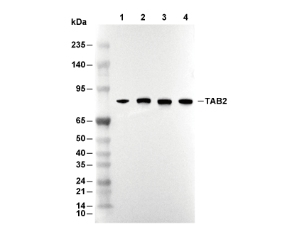 Lane 1: Hela, Lane 2: 293T, Lane 3: NIH/3T3, Lane 4: PC-12
Usage Information
| Dilution |
|---|
|
| Application |
|---|
| WB, IP |
| Reactivity |
|---|
| Human, Mouse, Rat |
| Source |
|---|
| Rabbit Monoclonal Antibody |
| Storage Buffer |
|---|
| PBS, pH 7.2+50% Glycerol+0.05% BSA+0.01% NaN3 |
| Storage (from the date of receipt) |
|---|
| -20°C (avoid freeze-thaw cycles), 2 years |
| Predicted MW Observed MW |
|---|
| 76 kDa 80 kDa,36 kDa |
| *Why do the predicted and actual molecular weights differ? The following reasons may explain differences between the predicted and actual protein molecular weight. Post-translational modifications(e.g., phosphorylation, glycosylation); Splice variants and isoforms; Relative charge; Multimerization. |
| Positive Control | Mouse brain tissue; Rat brain tissue; A549 cells; HEK-293 cells; Raji cells; HeLa cells; 293T cells; NIH/3T3 cells; PC-12 cells; THP-1 cells; RAW 264.7 cells; NR8383 cells |
|---|---|
| Negative Control | Jurkat cells |
Experimental Methods
| WB |
|---|
Experimental Protocol:
Sample preparation
1. Tissue: Lyse the tissue sample by adding an appropriate volume of ice-cold RIPA/NP-40 Lysis Buffer (containing Protease Inhibitor Cocktail),and homogenize the tissue at a low temperature or lyse it by sonication on ice, then incubate on ice for 30 minutes. 2. Adherent cell: Aspirate the culture medium and wash the cells with ice-cold PBS twice. Lyse the cells by adding an appropriate volume of RIPA/NP-40 Lysis Buffer (containing Protease Inhibitor Cocktail) , sonicate to lyse the cells, and incubate on ice for 30 minutes. 3. Suspension cell: Transfer the culture medium to a pre-cooled centrifuge tube. Centrifuge and aspirate the supernatant. Wash the cells with ice-cold PBS twice. Lyse the cells by adding an appropriate volume of RIPA/NP-40 Lysis Buffer (containing Protease Inhibitor Cocktail) , sonicate to lyse the cells, and incubate on ice for 30 minutes. 4. Place the lysate into a pre-cooled microcentrifuge tube. Centrifuge at 4°C for 15 min. Collect the supernatant;
5. Remove a small volume of lysate to determine the protein concentration;
6. Combine the lysate with protein loading buffer. Boil 20 µL sample under 95-100°C for 5 min. Centrifuge for 5 min after cool down on ice.
Electrophoretic separation
1. According to the concentration of extracted protein, load appropriate amount of protein sample and marker onto SDS-PAGE gels for electrophoresis. Recommended separating gel (lower gel) concentration: 10%. Reference Table for Selecting SDS-PAGE Separation Gel Concentrations 2. Power up 80V for 30 minutes. Then the power supply is adjusted (110 V~150 V), the Marker is observed, and the electrophoresis can be stopped when the indicator band of the predyed protein Marker where the protein is located is properly separated. (Note that the current should not be too large when electrophoresis, too large current (more than 150 mA) will cause the temperature to rise, affecting the result of running glue. If high currents cannot be avoided, an ice bath can be used to cool the bath.)
Transfer membrane
1. Take out the converter, soak the clip and consumables in the pre-cooled converter;
2. Activate PVDF membrane with methanol for 1 min and rinse with transfer buffer;
3. Install it in the order of "black edge of clip - sponge - filter paper - filter paper - glue -PVDF membrane - filter paper - filter paper - sponge - white edge of clip"; 4. The protein was electrotransferred to PVDF membrane. ( 0.45 µm PVDF membrane is recommended ) Reference Table for Selecting PVDF Membrane Pore Size Specifications Recommended conditions for wet transfer: 200 mA, 120 min. ( Note that the transfer conditions can be adjusted according to the protein size. For high-molecular-weight proteins, a higher current and longer transfer time are recommended. However, ensure that the transfer tank remains at a low temperature to prevent gel melting.)
Block
1. After electrotransfer, wash the film with TBST at room temperature for 5 minutes;
2. Incubate the film in the blocking solution for 1 hour at room temperature;
3. Wash the film with TBST for 3 times, 5 minutes each time.
Antibody incubation
1. Use 5% skim milk powder to prepare the primary antibody working liquid (recommended dilution ratio for primary antibody 1:1000), gently shake and incubate with the film at 4°C overnight; 2. Wash the film with TBST 3 times, 5 minutes each time;
3. Add the secondary antibody to the blocking solution and incubate with the film gently at room temperature for 1 hour;
4. After incubation, wash the film with TBST 3 times for 5 minutes each time.
Antibody staining
1. Add the prepared ECL luminescent substrate (or select other color developing substrate according to the second antibody) and mix evenly;
2. Incubate with the film for 1 minute, remove excess substrate (keep the film moist), wrap with plastic film, and expose in the imaging system. |
Biological Description
| Specificity |
|---|
| TAB2 Antibody (Rabbit mAb) [E6J10] detects endogenous levels of total TAB2 protein. |
| Subcellular Location |
|---|
| Cytoplasm, Endosome, Lysosome, Membrane |
| Uniprot ID |
|---|
| Q9NYJ8 |
| Clone |
|---|
| E6J10 |
| Synonym(s) |
|---|
| KIAA0733, MAP3K7IP2, TAB2, TAK1-binding protein 2, TGF-beta-activated kinase 1-binding protein 2, TAB-2 |
| Background |
|---|
| TAB2 (TAK1-binding protein 2/MAP3K7IP2) is a ubiquitin-binding adaptor of the TAK1–TAB scaffold family that links Lys63‑linked polyubiquitin signals generated downstream of receptors to activation of MAP3K7/TAK1 and divergent NF‑κB, JNK, and noncanonical Wnt pathways, while also exerting context‑specific roles in survival signaling and cardiac homeostasis. The protein contains an N‑terminal CUE/GLUE-like region and coiled-coil segments that mediate association with TAK1 and other partners, and a C‑terminal RanBP2-type zinc finger (NZF) domain that binds Lys63‑linked and mixed Lys63/Lys48‑linked polyubiquitin chains, unanchored or attached to substrates such as RIPK1 and RIPK2, creating a scaffold that positions TAK1 in proximity to ubiquitin-decorated receptor complexes and promotes TAK1 autophosphorylation. Upon stimulation by IL‑1 or TNF family ligands, TAB2 translocates from membranes to the cytosol and bridges TRAF6- or RIPK-containing ubiquitin platforms with TAK1–TAB1, enabling TAK1 activation and subsequent phosphorylation of the IKK core complex and MKKs that drive NF‑κB and JNK signaling, thereby coupling receptor-proximal ubiquitination to transcriptional responses that control inflammation, apoptosis, and proliferation. TAB2 also scaffolds TAK1 with Nemo-like kinase (NLK) in the Wnt cascade; in this setting TAB2-associated TAK1–NLK phosphorylates LEF1/TCF transcription factors and represses β‑catenin–dependent transcription, indicating that TAB2 can support noncanonical Wnt outputs that counterbalance canonical signaling during development and tissue homeostasis. Genetic and biochemical analyses show that TAB2 is not strictly required for all TAK1-dependent NF‑κB and MAPK activation, but performs additional functions in TNF signaling at the level of cytotoxic complex II, where its NZF-mediated binding to ubiquitinated components limits the abundance of apoptosis- and necroptosis-inducing complexes and maintains a pro‑survival checkpoint independently of TAK1 recruitment. In the myocardium, TAB2 has a critical role in preserving myocardial structure and function: TAB2 deficiency in mouse hearts promotes RIPK1-dependent apoptosis and necroptosis, amplifies inflammatory signaling, and leads to progressive dilated cardiomyopathy, showing that TAB2-dependent control of RIPK1-containing complexes is essential for myocardial homeostasis and remodeling under stress. Heterozygous TAB2 variants in families with syndromic congenital heart defects, adult-onset cardiomyopathy, and connective-tissue features, consistent with a dosage-sensitive requirement for TAB2-based signaling scaffolds during cardiac development and long-term ventricular integrity. TAB2 also interacts with nuclear receptor coregulators such as NCOR1 and estrogen receptor α in breast cancer cells, where defined central domains of TAB2 mediate dismissal of NCOR1 from antagonist-bound ERα on chromatin and contribute to tamoxifen resistance; disruption of this TAB2–ERα interface by competitive peptides restores antiproliferative responses to tamoxifen. |
| References |
|---|
|
Tech Support
Tel: +1-832-582-8158 Ext:3
If you have any other enquiries, please leave a message.
Products are for research use only. Not for human use. We do not sell to patients.
©Copyright 2013 Selleck Chemicals. All Rights Reserved.