-
 Australia
-
 Austria
-
 Belgium
-
 Brazil
-
 Canada
-
 China
China
-
 Czech Republic
-
 Denmark
-
 Finland
-
 France
-
 Germany
-
 Greece
-
 Hong Kong
-
 Hungary
-
 Iceland
-
 India
India
-
 Ireland
-
 Israel
-
 Italy
Italy
-
 Japan
Japan
-
 Korea
Korea
-
 Luxembourg
-
 Malaysia
-
 Netherlands
-
 New Zealand
-
 Norway
-
 Poland
-
 Qatar
Qatar
-
 Romania
-
 Saudi Arabia
-
 Singapore
-
 Spain
Spain
-
 Sweden
-
 Switzerland
-
 Taiwan
-
 Turkey
-
 United Kingdom
-
 United States
research use only
SPHK2 Antibody [A21H5]
Cat.No.: F6714
Application:
Reactivity:
-
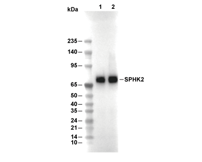 Lane 1: Raji, Lane 2: MOLT4
Usage Information
| Dilution |
|---|
|
| Application |
|---|
| WB, IP |
| Reactivity |
|---|
| Human |
| Source |
|---|
| Rabbit Monoclonal Antibody |
| Storage Buffer |
|---|
| PBS, pH 7.2+50% Glycerol+0.05% BSA+0.01% NaN3 |
| Storage (from the date of receipt) |
|---|
| -20°C (avoid freeze-thaw cycles), 2 years |
| Predicted MW |
|---|
| 75 kDa |
| Positive Control | Human kidney; Raji cells; MOLT-4 cells |
|---|---|
| Negative Control |
Experimental Methods
| WB |
|---|
Experimental Protocol:
Sample preparation
1. Tissue: Lyse the tissue sample by adding an appropriate volume of ice-cold RIPA/NP-40 Lysis Buffer (containing Protease Inhibitor Cocktail),and homogenize the tissue at a low temperature or lyse it by sonication on ice, then incubate on ice for 30 minutes. 2. Adherent cell: Aspirate the culture medium and wash the cells with ice-cold PBS twice. Lyse the cells by adding an appropriate volume of RIPA/NP-40 Lysis Buffer (containing Protease Inhibitor Cocktail), sonicate to lyse the cells, and incubate on ice for 30 minutes. 3. Suspension cell: Transfer the culture medium to a pre-cooled centrifuge tube. Centrifuge and aspirate the supernatant. Wash the cells with ice-cold PBS twice. Lyse the cells by adding an appropriate volume of RIPA/NP-40 Lysis Buffer (containing Protease Inhibitor Cocktail), sonicate to lyse the cells, and incubate on ice for 30 minutes. 4. Place the lysate into a pre-cooled microcentrifuge tube. Centrifuge at 4°C for 15 min. Collect the supernatant;
5. Remove a small volume of lysate to determine the protein concentration;
6. Combine the lysate with protein loading buffer. Boil 20 µL sample under 95-100°C for 5 min. Centrifuge for 5 min after cool down on ice.
Electrophoretic separation
1. According to the concentration of extracted protein, load appropriate amount of protein sample and marker onto SDS-PAGE gels for electrophoresis. Recommended separating gel (lower gel) concentration: 10%. Reference Table for Selecting SDS-PAGE Separation Gel Concentrations 2. Power up 80V for 30 minutes. Then the power supply is adjusted (110 V~150 V), the Marker is observed, and the electrophoresis can be stopped when the indicator band of the predyed protein Marker where the protein is located is properly separated. (Note that the current should not be too large when electrophoresis, too large current (more than 150 mA) will cause the temperature to rise, affecting the result of running glue. If high currents cannot be avoided, an ice bath can be used to cool the bath.)
Transfer membrane
1. Take out the converter, soak the clip and consumables in the pre-cooled converter;
2. Activate PVDF membrane with methanol for 1 min and rinse with transfer buffer;
3. Install it in the order of "black edge of clip - sponge - filter paper - filter paper - glue -PVDF membrane - filter paper - filter paper - sponge - white edge of clip"; 4. The protein was electrotransferred to PVDF membrane. ( 0.45 µm PVDF membrane is recommended ) Reference Table for Selecting PVDF Membrane Pore Size Specifications Recommended conditions for wet transfer: 200 mA, 120 min. ( Note that the transfer conditions can be adjusted according to the protein size. For high-molecular-weight proteins, a higher current and longer transfer time are recommended. However, ensure that the transfer tank remains at a low temperature to prevent gel melting.)
Block
1. After electrotransfer, wash the film with TBST at room temperature for 5 minutes;
2. Incubate the film in the blocking solution for 1 hour at room temperature;
3. Wash the film with TBST for 3 times, 5 minutes each time.
Antibody incubation
1. Use 5% skim milk powder to prepare the primary antibody working liquid (recommended dilution ratio for primary antibody 1:1000), gently shake and incubate with the film at 4°C overnight; 2. Wash the film with TBST 3 times, 5 minutes each time;
3. Add the secondary antibody to the blocking solution and incubate with the film gently at room temperature for 1 hour;
4. After incubation, wash the film with TBST 3 times for 5 minutes each time.
Antibody staining
1. Add the prepared ECL luminescent substrate (or select other color developing substrate according to the second antibody) and mix evenly;
2. Incubate with the film for 1 minute, remove excess substrate (keep the film moist), wrap with plastic film, and expose in the imaging system. |
Biological Description
| Specificity |
|---|
| SPHK2 Antibody [A21H5] detects endogenous levels of total SPHK2 protein. |
| Subcellular Location |
|---|
| Cytoplasm, Endoplasmic reticulum, Lysosome, Membrane, Mitochondrion, Mitochondrion inner membrane, Nucleus |
| Uniprot ID |
|---|
| Q9NRA0 |
| Clone |
|---|
| A21H5 |
| Synonym(s) |
|---|
| Sphingosine kinase 2; SPHK2 |
| Background |
|---|
| SPHK2 belongs to the sphingosine kinase family alongside SPHK1, catalyzing the ATP-dependent phosphorylation of sphingosine and related substrates to generate sphingosine-1-phosphate (S1P), a bioactive lipid mediator that governs intracellular signaling and intercellular communication through autocrine and paracrine actions. SPHK2 possesses a conserved kinase domain flanked by nuclear localization and export signals, enabling dynamic shuttling between cytoplasmic, nuclear, and organellar compartments such as the endoplasmic reticulum under stress conditions. Enzymatic activity centers on ATP-Mg binding to phosphorylate sphingoid bases, shifting the sphingolipid rheostat from pro-apoptotic ceramide and sphingosine toward pro-survival S1P, with substrate specificity extending to sphingadienine in lysosomal catabolism pathways. SPHK2-derived S1P associates with HDAC1/2 within repressor complexes to inhibit their deacetylase function, opening chromatin at promoters of cyclin genes and facilitating epigenetic activation of cell cycle progression alongside regulation of DNA damage responses. Cytoplasmic SPHK2 integrates signals from receptor tyrosine kinases and G-protein coupled receptors, undergoing ERK1-mediated phosphorylation at key serines and threonines to amplify S1P output during growth factor stimulation like EGF, thereby fueling PI3K/AKT/mTOR and MAPK/ERK cascades that drive migration, proliferation, and metabolic adaptation. SPHK2 further stabilizes telomerase reverse transcriptase (hTERT) by S1P binding, protecting telomeres from MKRN1-mediated ubiquitination and sustaining replicative potential in stem and progenitor cells. SPHK2 maintains mitochondrial respiration, telomere integrity, and autophagic flux in macrophages, where it prevents lipid droplet accumulation and foam cell formation during cholesterol efflux. In physiological contexts, SPHK2 enforces quiescence in hematopoietic and neural progenitors while promoting differentiation in response to cytokine gradients, with peak expression in brain and liver tissues reflecting roles in lipid quality control and preconditioning against ischemia. Dysregulation elevates SPHK2 in solid tumors and leukemias, where it confers chemoresistance via NF-κB/STAT3 activation and histone modifications, or paradoxically induces apoptosis when trafficked to stress granules. |
| References |
|---|
|
Tech Support
Tel: +1-832-582-8158 Ext:3
If you have any other enquiries, please leave a message.
Products are for research use only. Not for human use. We do not sell to patients.
©Copyright 2013 Selleck Chemicals. All Rights Reserved.