-
 Australia
-
 Austria
-
 Belgium
-
 Brazil
-
 Canada
-
 China
China
-
 Czech Republic
-
 Denmark
-
 Finland
-
 France
-
 Germany
-
 Greece
-
 Hong Kong
-
 Hungary
-
 Iceland
-
 India
India
-
 Ireland
-
 Israel
-
 Italy
Italy
-
 Japan
Japan
-
 Korea
Korea
-
 Luxembourg
-
 Malaysia
-
 Netherlands
-
 New Zealand
-
 Norway
-
 Poland
-
 Qatar
Qatar
-
 Romania
-
 Saudi Arabia
-
 Singapore
-
 Spain
Spain
-
 Sweden
-
 Switzerland
-
 Taiwan
-
 Turkey
-
 United Kingdom
-
 United States
research use only
SHOC2 Antibody [H23M19]
Cat.No.: F5330
Application:
Reactivity:
-
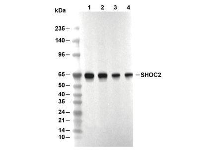 Lane 1: T47D, Lane 2: HCT116, Lane 3: HT29, Lane 4: C6
Usage Information
| Dilution |
|---|
|
| Application |
|---|
| WB |
| Reactivity |
|---|
| Human, Mouse, Rat, Monkey |
| Source |
|---|
| Rabbit Monoclonal Antibody |
| Storage Buffer |
|---|
| PBS, pH 7.2+50% Glycerol+0.05% BSA+0.01% NaN3 |
| Storage (from the date of receipt) |
|---|
| -20°C (avoid freeze-thaw cycles), 2 years |
| Predicted MW |
|---|
| 64 kDa |
| Positive Control | 786-O cells; TK-10 cells; KM12 cells; LNCaP cells; T-47D cells; HCT 116 cells; HT-29 cells; A549 cells; NIH/3T3 cells; C6 cells; COS-7 cells |
|---|---|
| Negative Control |
Experimental Methods
| WB |
|---|
Experimental Protocol:
Sample preparation
1. Tissue: Lyse the tissue sample by adding an appropriate volume of ice-cold RIPA/NP-40 Lysis Buffer (containing Protease Inhibitor Cocktail),and homogenize the tissue at a low temperature or lyse it by sonication on ice, then incubate on ice for 30 minutes. 2. Adherent cell: Aspirate the culture medium and wash the cells with ice-cold PBS twice. Lyse the cells by adding an appropriate volume of RIPA/NP-40 Lysis Buffer (containing Protease Inhibitor Cocktail), sonicate to lyse the cells, and incubate on ice for 30 minutes. 3. Suspension cell: Transfer the culture medium to a pre-cooled centrifuge tube. Centrifuge and aspirate the supernatant. Wash the cells with ice-cold PBS twice. Lyse the cells by adding an appropriate volume of RIPA/NP-40 Lysis Buffer (containing Protease Inhibitor Cocktail), sonicate to lyse the cells, and incubate on ice for 30 minutes. 4. Place the lysate into a pre-cooled microcentrifuge tube. Centrifuge at 4°C for 15 min. Collect the supernatant;
5. Remove a small volume of lysate to determine the protein concentration;
6. Combine the lysate with protein loading buffer. Boil 20 µL sample under 95-100°C for 5 min. Centrifuge for 5 min after cool down on ice.
Electrophoretic separation
1. According to the concentration of extracted protein, load appropriate amount of protein sample and marker onto SDS-PAGE gels for electrophoresis. Recommended separating gel (lower gel) concentration: 10%. Reference Table for Selecting SDS-PAGE Separation Gel Concentrations 2. Power up 80V for 30 minutes. Then the power supply is adjusted (110 V~150 V), the Marker is observed, and the electrophoresis can be stopped when the indicator band of the predyed protein Marker where the protein is located is properly separated. (Note that the current should not be too large when electrophoresis, too large current (more than 150 mA) will cause the temperature to rise, affecting the result of running glue. If high currents cannot be avoided, an ice bath can be used to cool the bath.)
Transfer membrane
1. Take out the converter, soak the clip and consumables in the pre-cooled converter;
2. Activate PVDF membrane with methanol for 1 min and rinse with transfer buffer;
3. Install it in the order of "black edge of clip - sponge - filter paper - filter paper - glue -PVDF membrane - filter paper - filter paper - sponge - white edge of clip"; 4. The protein was electrotransferred to PVDF membrane. ( 0.45 µm PVDF membrane is recommended ) Reference Table for Selecting PVDF Membrane Pore Size Specifications Recommended conditions for wet transfer: 200 mA, 120 min. ( Note that the transfer conditions can be adjusted according to the protein size. For high-molecular-weight proteins, a higher current and longer transfer time are recommended. However, ensure that the transfer tank remains at a low temperature to prevent gel melting.)
Block
1. After electrotransfer, wash the film with TBST at room temperature for 5 minutes;
2. Incubate the film in the blocking solution for 1 hour at room temperature;
3. Wash the film with TBST for 3 times, 5 minutes each time.
Antibody incubation
1. Use 5% skim milk powder to prepare the primary antibody working liquid (recommended dilution ratio for primary antibody 1:1000), gently shake and incubate with the film at 4°C overnight; 2. Wash the film with TBST 3 times, 5 minutes each time;
3. Add the secondary antibody to the blocking solution and incubate with the film gently at room temperature for 1 hour;
4. After incubation, wash the film with TBST 3 times for 5 minutes each time.
Antibody staining
1. Add the prepared ECL luminescent substrate (or select other color developing substrate according to the second antibody) and mix evenly;
2. Incubate with the film for 1 minute, remove excess substrate (keep the film moist), wrap with plastic film, and expose in the imaging system. |
Biological Description
| Specificity |
|---|
| SHOC2 Antibody [H23M19] detects endogenous levels of total SHOC2 protein. |
| Subcellular Location |
|---|
| Cytoplasm, Nucleus |
| Uniprot ID |
|---|
| Q9UQ13 |
| Clone |
|---|
| H23M19 |
| Synonym(s) |
|---|
| Leucine-rich repeat protein SHOC-2; Ras-binding protein Sur-8 homolog; SHOC2 |
| Background |
|---|
| SHOC2 is a leucine‑rich repeat (LRR)‑containing scaffold protein that positively regulates RAS‑MAPK signaling and intersects with the PI3K/Akt axis, functioning as a modular platform that coordinates the assembly and dynamics of specific signaling complexes. SHOC2 folds into a concave LRR‑rich domain that mediates multiple protein–protein interactions, enabling its recruitment to activated RAS‑GTP and to the catalytic subunit of protein phosphatase 1 (PP1), while its N‑terminal region participates in competitive membrane association and regulatory switches. SHOC2 forms a ternary complex with a regulatory PP1 subunit and the GTP‑bound small GTPase M‑RAS, and this complex serves as a dedicated accelerator of RAF–MEK–ERK signaling, promoting RAF dimerization and MEK/ERK phosphorylation downstream of receptor‑tyrosine‑kinase‑driven growth‑factor inputs. SHOC2 engages the p110α catalytic subunit of PI3K, thereby linking RAS‑MAPK activation to PI3K/Akt stimulation and broadening the range of downstream transcriptional and metabolic outputs that depend on coordinated ERK and Akt signaling. This dual engagement of ERK and PI3K/Akt pathways underlies SHOC2’s role in promoting cell motility, cytoskeletal remodeling, and invasive behavior in epithelial and cancer cell contexts, where its expression supports processes that increase migratory capacity and metastatic potential. A germline SHOC2 mutation that introduces an N‑terminal myristoylation motif causes aberrant constitutive membrane targeting of the protein, leading to sustained, growth‑factor‑independent enhancement of RAF–ERK signaling and underpinning the hyperactive RAS–MAPK phenotype in Noonan‑like syndrome with loose anagen hair. |
| References |
|---|
|
Tech Support
Tel: +1-832-582-8158 Ext:3
If you have any other enquiries, please leave a message.
Products are for research use only. Not for human use. We do not sell to patients.
©Copyright 2013 Selleck Chemicals. All Rights Reserved.