-
 Australia
-
 Austria
-
 Belgium
-
 Brazil
-
 Canada
-
 China
China
-
 Czech Republic
-
 Denmark
-
 Finland
-
 France
-
 Germany
-
 Greece
-
 Hong Kong
-
 Hungary
-
 Iceland
-
 India
India
-
 Ireland
-
 Israel
-
 Italy
Italy
-
 Japan
Japan
-
 Korea
Korea
-
 Luxembourg
-
 Malaysia
-
 Netherlands
-
 New Zealand
-
 Norway
-
 Poland
-
 Qatar
Qatar
-
 Romania
-
 Saudi Arabia
-
 Singapore
-
 Spain
Spain
-
 Sweden
-
 Switzerland
-
 Taiwan
-
 Turkey
-
 United Kingdom
-
 United States
research use only
PTBP1 Antibody [L20G7]
Cat.No.: F5005
Application:
Reactivity:
-
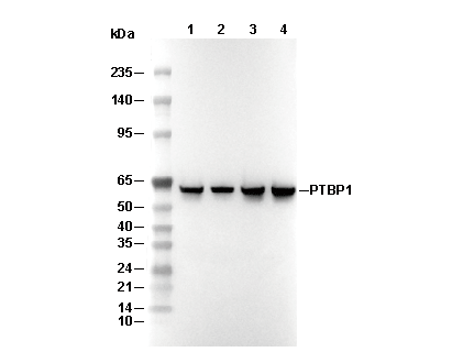 Lane 1: Hela, Lane 2: NIH/3T3, Lane 3: C6, Lane 4: COS-7
Usage Information
| Dilution |
|---|
|
| Application |
|---|
| WB, IP, ChIP |
| Reactivity |
|---|
| Human, Mouse, Rat, Monkey |
| Source |
|---|
| Rabbit Monoclonal Antibody |
| Storage Buffer |
|---|
| PBS, pH 7.2+50% Glycerol+0.05% BSA+0.01% NaN3 |
| Storage (from the date of receipt) |
|---|
| -20°C (avoid freeze-thaw cycles), 2 years |
| Predicted MW |
|---|
| 57 kDa, 59 kDa |
| Positive Control | K-562 cells; MCF7 cells; HeLa cells; NIH/3T3 cells; C6 cells; COS-7 cells |
|---|---|
| Negative Control |
Experimental Methods
| WB |
|---|
Experimental Protocol:
Sample preparation
1. Tissue: Lyse the tissue sample by adding an appropriate volume of ice-cold RIPA/Nuclear Lysis Buffer (containing Protease Inhibitor Cocktail),and homogenize the tissue at a low temperature or lyse it by sonication on ice, then incubate on ice for 30 minutes. 2. Adherent cell: Aspirate the culture medium and wash the cells with ice-cold PBS twice. Lyse the cells by adding an appropriate volume of RIPA/Nuclear Lysis Buffer (containing Protease Inhibitor Cocktail), sonicate to lyse the cells, and incubate on ice for 30 minutes. 3. Suspension cell: Transfer the culture medium to a pre-cooled centrifuge tube. Centrifuge and aspirate the supernatant. Wash the cells with ice-cold PBS twice. Lyse the cells by adding an appropriate volume of RIPA/Nuclear Lysis Buffer (containing Protease Inhibitor Cocktail), sonicate to lyse the cells, and incubate on ice for 30 minutes. 4. Place the lysate into a pre-cooled microcentrifuge tube. Centrifuge at 4°C for 15 min. Collect the supernatant;
5. Remove a small volume of lysate to determine the protein concentration;
6. Combine the lysate with protein loading buffer. Boil 20 µL sample under 95-100°C for 5 min. Centrifuge for 5 min after cool down on ice.
Electrophoretic separation
1. According to the concentration of extracted protein, load appropriate amount of protein sample and marker onto SDS-PAGE gels for electrophoresis. Recommended separating gel (lower gel) concentration: 10%. Reference Table for Selecting SDS-PAGE Separation Gel Concentrations 2. Power up 80V for 30 minutes. Then the power supply is adjusted (110 V~150 V), the Marker is observed, and the electrophoresis can be stopped when the indicator band of the predyed protein Marker where the protein is located is properly separated. (Note that the current should not be too large when electrophoresis, too large current (more than 150 mA) will cause the temperature to rise, affecting the result of running glue. If high currents cannot be avoided, an ice bath can be used to cool the bath.)
Transfer membrane
1. Take out the converter, soak the clip and consumables in the pre-cooled converter;
2. Activate PVDF membrane with methanol for 1 min and rinse with transfer buffer;
3. Install it in the order of "black edge of clip - sponge - filter paper - filter paper - glue -PVDF membrane - filter paper - filter paper - sponge - white edge of clip"; 4. The protein was electrotransferred to PVDF membrane. ( 0.45 µm PVDF membrane is recommended ) Reference Table for Selecting PVDF Membrane Pore Size Specifications Recommended conditions for wet transfer: 200 mA, 120 min. ( Note that the transfer conditions can be adjusted according to the protein size. For high-molecular-weight proteins, a higher current and longer transfer time are recommended. However, ensure that the transfer tank remains at a low temperature to prevent gel melting.)
Block
1. After electrotransfer, wash the film with TBST at room temperature for 5 minutes;
2. Incubate the film in the blocking solution for 1 hour at room temperature;
3. Wash the film with TBST for 3 times, 5 minutes each time.
Antibody incubation
1. Use 5% skim milk powder to prepare the primary antibody working liquid (recommended dilution ratio for primary antibody 1:1000), gently shake and incubate with the film at 4°C overnight; 2. Wash the film with TBST 3 times, 5 minutes each time;
3. Add the secondary antibody to the blocking solution and incubate with the film gently at room temperature for 1 hour;
4. After incubation, wash the film with TBST 3 times for 5 minutes each time.
Antibody staining
1. Add the prepared ECL luminescent substrate (or select other color developing substrate according to the second antibody) and mix evenly;
2. Incubate with the film for 1 minute, remove excess substrate (keep the film moist), wrap with plastic film, and expose in the imaging system. |
Biological Description
| Specificity |
|---|
| PTBP1 Antibody [L20G7] detects endogenous levels of total PTBP1 protein. |
| Subcellular Location |
|---|
| Nucleus |
| Uniprot ID |
|---|
| P26599-1 |
| Clone |
|---|
| L20G7 |
| Synonym(s) |
|---|
| Polypyrimidine tract-binding protein 1; PTB; 57 kDa RNA-binding protein PPTB-1; Heterogeneous nuclear ribonucleoprotein I (hnRNP I); PTBP1; PTB |
| Background |
|---|
| PTBP1 (Polypyrimidine Tract Binding Protein 1) is a ubiquitously expressed RNA-binding protein of the PTB family that silences alternative exon splicing by binding polypyrimidine tracts on pre-mRNA, regulating neural differentiation, cell proliferation, and oncogenic pathways. It features an N-terminal NLS/NES and four heterogeneous RRMs (RRM1-4) in "beads-on-a-string" configuration with flexible linkers (42 aa after RRM1, 58-84 aa after RRM2), where each RRM's αβ-sandwich binds U/C-rich sequences via distinct hydrophobic pockets (RRM1: L89/L91/F98/H133/L136; RRM2: I214/F216/L225/S258). PTBP1's primary function is splicing repression of exons like PKM (favoring cancer-associated PKM2), with RRM3-4 forming a rigid didomain that manipulates RNA structure while RRM1/2 move independently for specificity; PTBP1 outcompetes U2AF65 and recruits cofactors Raver1/Matrin3 for stronger repression than neural paralog PTBP2. It maintains epithelial identity, blocks neuronal genes in non-neurals (PTBP1→PTBP2 switch enables neurogenesis), and drives cancer via PKM2/CDK6/EXOC7 exon inclusion promoting glycolysis/proliferation/invasion; overexpressed in cancers correlating with poor prognosis. Disease relevance includes glioblastoma (PTBP1 suppression induces proneural-to-mesenchymal transition) and therapeutic targeting via antisense oligonucleotides to reverse oncogenic splicing. |
| References |
|---|
|
Tech Support
Tel: +1-832-582-8158 Ext:3
If you have any other enquiries, please leave a message.
Products are for research use only. Not for human use. We do not sell to patients.
©Copyright 2013 Selleck Chemicals. All Rights Reserved.