-
 Australia
-
 Austria
-
 Belgium
-
 Brazil
-
 Canada
-
 China
China
-
 Czech Republic
-
 Denmark
-
 Finland
-
 France
-
 Germany
-
 Greece
-
 Hong Kong
-
 Hungary
-
 Iceland
-
 India
India
-
 Ireland
-
 Israel
-
 Italy
Italy
-
 Japan
Japan
-
 Korea
Korea
-
 Luxembourg
-
 Malaysia
-
 Netherlands
-
 New Zealand
-
 Norway
-
 Poland
-
 Qatar
Qatar
-
 Romania
-
 Saudi Arabia
-
 Singapore
-
 Spain
Spain
-
 Sweden
-
 Switzerland
-
 Taiwan
-
 Turkey
-
 United Kingdom
-
 United States
research use only
Polycystin 1/PC1 Antibody [B20F13]
Cat.No.: F2579
Application:
Reactivity:
-
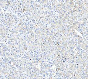 Immunohistochemical analysis of formalin fixed paraffin embedded human liver tissue with F2579 at 1:200 dilution.
Usage Information
| Dilution |
|---|
|
| Application |
|---|
| IHC |
| Reactivity |
|---|
| Human |
| Source |
|---|
| Mouse Monoclonal Antibody |
| Storage Buffer |
|---|
| PBS, pH 7.2+50% Glycerol+0.05% BSA+0.01% NaN3 |
| Storage (from the date of receipt) |
|---|
| -20°C (avoid freeze-thaw cycles), 2 years |
| Positive Control | Normal Human Bone Marrow; Human Normal Liver; Normal human kidney tissue |
|---|---|
| Negative Control |
Experimental Methods
| IHC |
|---|
Experimental Protocol:
Deparaffinization/Rehydration
1. Deparaffinize/hydrate sections:
2. Incubate sections in three washes of xylene for 5 min each.
3. Incubate sections in two washes of 100% ethanol for 10 min each.
4. Incubate sections in two washes of 95% ethanol for 10 min each.
5. Wash sections two times in dH2O for 5 min each.
6.Antigen retrieval: For Citrate: Heat slides in a microwave submersed in 1X citrate unmasking solution until boiling is initiated; continue with 10 min at a sub-boiling temperature (95°-98°C). Cool slides on bench top for 30 min.
Staining
1. Wash sections in dH2O three times for 5 min each.
2. Incubate sections in 3% hydrogen peroxide for 10 min.
3. Wash sections in dH2O two times for 5 min each.
4. Wash sections in wash buffer for 5 min.
5. Block each section with 100–400 µl of blocking solution for 1 hr at room temperature.
6. Remove blocking solution and add 100–400 µl primary antibody diluent in to each section. Incubate overnight at 4°C.
7. Remove antibody solution and wash sections with wash buffer three times for 5 min each.
8. Cover section with 1–3 drops HRPas needed. Incubate in a humidified chamber for 30 min at room temperature.
9. Wash sections three times with wash buffer for 5 min each.
10. Add DAB Chromogen Concentrate to DAB Diluent and mix well before use.
11. Apply 100–400 µl DAB to each section and monitor closely. 1–10 min generally provides an acceptable staining intensity.
12. Immerse slides in dH2O.
13. If desired, counterstain sections with hematoxylin.
14. Wash sections in dH2O two times for 5 min each.
15. Dehydrate sections: Incubate sections in 95% ethanol two times for 10 sec each; Repeat in 100% ethanol, incubating sections two times for 10 sec each; Repeat in xylene, incubating sections two times for 10 sec each.
16. Mount sections with coverslips and mounting medium.
|
Biological Description
| Specificity |
|---|
| Polycystin 1/PC1 Antibody [B20F13] detects endogenous levels of total Polycystin 1/PC1 protein. |
| Subcellular Location |
|---|
| Cell membrane, Cell projection, Cilium, Endoplasmic reticulum, Golgi apparatus, Membrane, Secreted |
| Uniprot ID |
|---|
| P98161 |
| Clone |
|---|
| B20F13 |
| Synonym(s) |
|---|
| Polycystin‑1; PC1; Autosomal dominant polycystic kidney disease 1 protein; PKD1 |
| Background |
|---|
| Polycystin 1 (PC1), encoded by the PKD1 gene, is a large transmembrane protein that functions as a mechanosensitive receptor in epithelial cells of renal tubules, liver, and pancreas, where it regulates cell–matrix adhesion, lumen architecture, and mechanotransduction. Its extended extracellular domain integrates multiple structural motifs that engage mechanical strain and ligand dependent cues at the cell surface and couples through the membrane to polycystin 2 (PC2), assembling a calcium permeable channel complex that localizes to primary cilia and apical membranes and converts luminal flow and shear stress into graded intracellular calcium signals. These PC1–PC2 dependent calcium fluxes intersect with calcineurin, mTORC1, ERK, and JAK–STAT pathways to modulate cell proliferation, differentiation, fluid secretion, and cytoskeletal remodeling, while PC1 simultaneously engages Wnt–planar cell polarity and integrin linked signaling to coordinate planar orientation of mitotic spindles, apicobasal polarization, and extracellular matrix organization across the tubular epithelium. In autosomal dominant polycystic kidney disease, germline or somatic inactivating PKD1 mutations impair PC1–PC2 complex formation, trafficking, or channel gating, leading to sustained suppression of calcium dependent growth inhibitory signals and aberrant activation of mTORC1 and proliferative kinase cascades that drive epithelial hyperplasia and misoriented divisions. This altered signaling environment promotes progressive tubular dilation, mislocalized fluid secretion, and matrix remodeling, resulting in cyst expansion in kidneys and liver, and analogous PC2 lesions produce a comparable phenotype, demonstrating the essential interdependence of both polycystins in maintaining tubular integrity, such that PC1 dependent signaling is ultimately dysregulated in the cystic epithelium. |
| References |
|---|
|
Tech Support
Tel: +1-832-582-8158 Ext:3
If you have any other enquiries, please leave a message.
Products are for research use only. Not for human use. We do not sell to patients.
©Copyright 2013 Selleck Chemicals. All Rights Reserved.