-
 Australia
-
 Austria
-
 Belgium
-
 Brazil
-
 Canada
-
 China
China
-
 Czech Republic
-
 Denmark
-
 Finland
-
 France
-
 Germany
-
 Greece
-
 Hong Kong
-
 Hungary
-
 Iceland
-
 India
India
-
 Ireland
-
 Israel
-
 Italy
Italy
-
 Japan
Japan
-
 Korea
Korea
-
 Luxembourg
-
 Malaysia
-
 Netherlands
-
 New Zealand
-
 Norway
-
 Poland
-
 Qatar
Qatar
-
 Romania
-
 Saudi Arabia
-
 Singapore
-
 Spain
Spain
-
 Sweden
-
 Switzerland
-
 Taiwan
-
 Turkey
-
 United Kingdom
-
 United States
research use only
Phospho-RIP3 (Ser232) Antibody (Rabbit mAb) [E19K17]
CatNo: F1682
Application:
Reactivity:
-
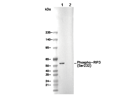 Lane 1: L-929, Lane 2: L-929 (phosphatase-treated)
Usage Information
| Dilution |
|---|
|
| Application |
|---|
| WB, ELISA |
| Reactivity |
|---|
| Mouse |
| Source |
|---|
| Rabbit Monoclonal Antibody |
| Storage Buffer |
|---|
| PBS, pH 7.2+50% Glycerol+0.05% BSA+0.01% NaN3 |
| Storage (from the date of receipt) |
|---|
| -20°C (avoid freeze-thaw cycles), 2 years |
| Predicted MW |
|---|
| 53 kDa |
| Positive Control | L-929 cell (treated with 20 ng/ml TNF-a, 100 nM Smac mimetic and 20 µM z-VAD for 6-8 hours), TSE cell (treated with 20 ng/ml TNF alpha,100 nM Smac mimetic, and 20 µM z-VAD for 12 h) |
|---|---|
| Negative Control |
Experimental Methods
| WB |
|---|
Experimental Protocol:
Sample preparation
1. Tissue: Lyse the tissue sample by adding an appropriate volume of ice-cold RIPA/Nuclear Lysis Buffer (containing Protease Inhibitor Cocktail, Phosphatase Inhibitor Cocktail),and homogenize the tissue at a low temperature or lyse it by sonication on ice, then incubate on ice for 30 minutes. 2. Adherent cell: Aspirate the culture medium and wash the cells with ice-cold PBS twice. Lyse the cells by adding an appropriate volume of RIPA/Nuclear Lysis Buffer (containing Protease Inhibitor Cocktail, Phosphatase Inhibitor Cocktail), sonicate to lyse the cells, and incubate on ice for 30 minutes. 3. Suspension cell: Transfer the culture medium to a pre-cooled centrifuge tube. Centrifuge and aspirate the supernatant. Wash the cells with ice-cold PBS twice. Lyse the cells by adding an appropriate volume of RIPA/Nuclear Lysis Buffer (containing Protease Inhibitor Cocktail, Phosphatase Inhibitor Cocktail), sonicate to lyse the cells, and incubate on ice for 30 minutes. 4. Place the lysate into a pre-cooled microcentrifuge tube. Centrifuge at 4°C for 15 min. Collect the supernatant;
5. Remove a small volume of lysate to determine the protein concentration;
6. Combine the lysate with protein loading buffer. Boil 20 µL sample under 95-100°C for 5 min. Centrifuge for 5 min after cool down on ice.
Electrophoretic separation
1. According to the concentration of extracted protein, load appropriate amount of protein sample and marker onto SDS-PAGE gels for electrophoresis. Recommended separating gel (lower gel) concentration: 10%. Reference Table for Selecting SDS-PAGE Separation Gel Concentrations 2. Power up 80V for 30 minutes. Then the power supply is adjusted (110 V~150 V), the Marker is observed, and the electrophoresis can be stopped when the indicator band of the predyed protein Marker where the protein is located is properly separated. (Note that the current should not be too large when electrophoresis, too large current (more than 150 mA) will cause the temperature to rise, affecting the result of running glue. If high currents cannot be avoided, an ice bath can be used to cool the bath.)
Transfer membrane
1. Take out the converter, soak the clip and consumables in the pre-cooled converter;
2. Activate PVDF membrane with methanol for 1 min and rinse with transfer buffer;
3. Install it in the order of "black edge of clip - sponge - filter paper - filter paper - glue -PVDF membrane - filter paper - filter paper - sponge - white edge of clip"; 4. The protein was electrotransferred to PVDF membrane. ( 0.45 µm PVDF membrane is recommended ) Reference Table for Selecting PVDF Membrane Pore Size Specifications Recommended conditions for wet transfer: 200 mA, 120 min. ( Note that the transfer conditions can be adjusted according to the protein size. For high-molecular-weight proteins, a higher current and longer transfer time are recommended. However, ensure that the transfer tank remains at a low temperature to prevent gel melting.)
Block
1. After electrotransfer, wash the film with TBST at room temperature for 5 minutes;
2. Incubate the film in the blocking solution ( recommending 5% BSA solution)
for 1 hour at room temperature;
3. Wash the film with TBST for 3 times, 5 minutes each time.
Antibody incubation
1. Use 5% skim milk powder to prepare the primary antibody working liquid (recommended dilution ratio for primary antibody 1:1000), gently shake and incubate with the film at 4°C overnight; 2. Wash the film with TBST 3 times, 5 minutes each time;
3. Add the secondary antibody to the blocking solution and incubate with the film gently at room temperature for 1 hour;
4. After incubation, wash the film with TBST 3 times for 5 minutes each time.
Antibody staining
1. Add the prepared ECL luminescent substrate (or select other color developing substrate according to the second antibody) and mix evenly;
2. Incubate with the film for 1 minute, remove excess substrate (keep the film moist), wrap with plastic film, and expose in the imaging system. |
Biological Description
| Specificity |
|---|
Phospho-RIP3 (Ser232) Antibody (Rabbit mAb) [E19K17] recognizes endogenous levels of total RIP3 protein only when phosphorylated at Ser232 protein |
| Subcellular Location |
|---|
| Cytoplasm, Nucleus |
| Uniprot ID |
|---|
| Q9QZL0 |
| Clone |
|---|
| E19K17 |
| Synonym(s) |
|---|
| Rip3, Ripk3, Receptor-interacting serine/threonine-protein kinase 3, RIP-like protein kinase 3, Receptor-interacting protein 3, RIP-3, mRIP3 |
| Background |
|---|
| The receptor-interacting protein (RIP) family of serine/threonine kinases—including RIP1, RIP2, RIP3, and RIP4—plays a crucial role in modulating cellular stress responses. These kinases are involved in initiating pro-survival and inflammatory signaling, primarily through NF-κB activation, as well as in promoting cell death pathways such as apoptosis. Among them, receptor-interacting protein kinase 3 (RIPK3 or RIP3) is a central player in necroptosis, a regulated form of necrotic cell death. RIP3 participates in signaling cascades initiated by tumor necrosis factor (TNF), where it associates with RIP1 and the TNF receptor complex to mediate both NF-κB activation and apoptosis. A key event in necroptosis is the interaction between RIP1 and RIP3, which forms the basis of the necrosome—a multiprotein complex that facilitates programmed necrosis. This necrotic-like cell death is typically triggered by TNF signaling in the presence of caspase inhibition. In humans, phosphorylation of RIP3 at Serine 227 is critical for its binding to mixed lineage kinase domain-like protein (MLKL), enabling necrosome assembly. In mice, TNF stimulation induces phosphorylation at Threonine 231 and Serine 232 on RIP3, which is necessary for its interaction with mouse MLKL. Notably, Ser-232 in murine RIP3 corresponds to Ser-227 in the human protein. Additionally, RIP1 and RIP3 contribute to necrosome oligomerization, which promotes the formation of amyloid-like signaling structures essential for executing necroptosis. |
| References |
|---|
|
Tech Support
Tel: +1-832-582-8158 Ext:3
If you have any other enquiries, please leave a message.
Products are for research use only. Not for human use. We do not sell to patients.
©Copyright 2013 Selleck Chemicals. All Rights Reserved.