-
 Australia
-
 Austria
-
 Belgium
-
 Brazil
-
 Canada
-
 China
China
-
 Czech Republic
-
 Denmark
-
 Finland
-
 France
-
 Germany
-
 Greece
-
 Hong Kong
-
 Hungary
-
 Iceland
-
 India
India
-
 Ireland
-
 Israel
-
 Italy
Italy
-
 Japan
Japan
-
 Korea
Korea
-
 Luxembourg
-
 Malaysia
-
 Netherlands
-
 New Zealand
-
 Norway
-
 Poland
-
 Qatar
Qatar
-
 Romania
-
 Saudi Arabia
-
 Singapore
-
 Spain
Spain
-
 Sweden
-
 Switzerland
-
 Taiwan
-
 Turkey
-
 United Kingdom
-
 United States
research use only
Phospho-RAB8A (Thr72) Antibody [A18L3]
Cat.No.: F2499
Application:
Reactivity:
-
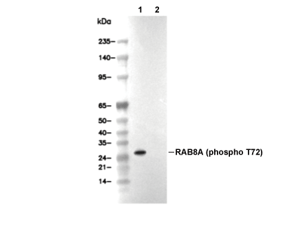 Lane 1: MEF, Lane 2: MEF (MLi-2, 100 nM, 90 min)
Experiment Essentials
WB
Recommended wet transfer conditions: 200 mA, 60 min.
Recommended wet transfer conditions: 200 mA, 60 min.
Usage Information
| Dilution |
|---|
|
| Application |
|---|
| WB |
| Reactivity |
|---|
| Mouse |
| Source |
|---|
| Rabbit Monoclonal Antibody |
| Storage Buffer |
|---|
| PBS, pH 7.2+50% Glycerol+0.05% BSA+0.01% NaN3 |
| Storage (from the date of receipt) |
|---|
| -20°C (avoid freeze-thaw cycles), 2 years |
| Predicted MW |
|---|
| 24 kDa |
| Positive Control | MEF cells |
|---|---|
| Negative Control |
Experimental Methods
| WB |
|---|
Experimental Protocol:
Sample preparation
1. Tissue: Lyse the tissue sample by adding an appropriate volume of ice-cold RIPA/NP-40 Lysis Buffer (containing Protease Inhibitor Cocktail, Phosphatase Inhibitor Cocktail),and homogenize the tissue at a low temperature. 2. Adherent cell: Aspirate the culture medium and wash the cells with ice-cold PBS twice. Lyse the cells by adding an appropriate volume of RIPA/NP-40 Lysis Buffer (containing Protease Inhibitor Cocktail, Phosphatase Inhibitor Cocktail) and put the sample on ice for 5 min. 3. Suspension cell: Transfer the culture medium to a pre-cooled centrifuge tube. Centrifuge and aspirate the supernatant. Wash the cells with ice-cold PBS twice. Lyse the cells by adding an appropriate volume of RIPA/NP-40 Lysis Buffer (containing Protease Inhibitor Cocktail, Phosphatase Inhibitor Cocktail) and put the sample on ice for 5 min. 4. Place the lysate into a pre-cooled microcentrifuge tube. Centrifuge at 4°C for 15 min. Collect the supernatant;
5. Remove a small volume of lysate to determine the protein concentration;
6. Combine the lysate with protein loading buffer. Boil 20 µL sample under 95-100°C for 5 min. Centrifuge for 5 min after cool down on ice.
Electrophoretic separation
1. According to the concentration of extracted protein, load appropriate amount of protein sample and marker onto SDS-PAGE gels for electrophoresis. Recommended separating gel (lower gel) concentration: 10%. Reference Table for Selecting SDS-PAGE Separation Gel Concentrations 2. Power up 80V for 30 minutes. Then the power supply is adjusted (110 V~150 V), the Marker is observed, and the electrophoresis can be stopped when the indicator band of the predyed protein Marker where the protein is located is properly separated. (Note that the current should not be too large when electrophoresis, too large current (more than 150 mA) will cause the temperature to rise, affecting the result of running glue. If high currents cannot be avoided, an ice bath can be used to cool the bath.)
Transfer membrane
1. Take out the converter, soak the clip and consumables in the pre-cooled converter;
2. Activate PVDF membrane with methanol for 1 min and rinse with transfer buffer;
3. Install it in the order of "black edge of clip - sponge - filter paper - filter paper - glue -PVDF membrane - filter paper - filter paper - sponge - white edge of clip"; 4. The protein was electrotransferred to PVDF membrane. ( 0.45 µm PVDF membrane is recommended ) Reference Table for Selecting PVDF Membrane Pore Size Specifications Recommended conditions for wet transfer: 200 mA, 60 min. ( Note that the transfer conditions can be adjusted according to the protein size. For high-molecular-weight proteins, a higher current and longer transfer time are recommended. However, ensure that the transfer tank remains at a low temperature to prevent gel melting.)
Block
1. After electrotransfer, wash the film with TBST at room temperature for 5 minutes;
2. Incubate the film in the blocking solution ( recommending 5% BSA solution)
for 1 hour at room temperature;
3. Wash the film with TBST for 3 times, 5 minutes each time.
Antibody incubation
1. Use 5% skim milk powder to prepare the primary antibody working liquid (recommended dilution ratio for primary antibody 1:1000), gently shake and incubate with the film at 4°C overnight; 2. Wash the film with TBST 3 times, 5 minutes each time;
3. Add the secondary antibody to the blocking solution and incubate with the film gently at room temperature for 1 hour;
4. After incubation, wash the film with TBST 3 times for 5 minutes each time.
Antibody staining
1. Add the prepared ECL luminescent substrate (or select other color developing substrate according to the second antibody) and mix evenly;
2. Incubate with the film for 1 minute, remove excess substrate (keep the film moist), wrap with plastic film, and expose in the imaging system. |
Biological Description
| Specificity |
|---|
| Phospho-RAB8A (Thr72) Antibody [A18L3] detects endogenous levels of total RAB8A protein only when it is phosphorylated at Thr72. |
| Subcellular Location |
|---|
| Cell membrane, Cell projection, Cilium, Cytoplasm, Cytoplasmic vesicle, Cytoskeleton, Endosome, Golgi apparatus, Lysosome |
| Uniprot ID |
|---|
| P61006 |
| Clone |
|---|
| A18L3 |
| Synonym(s) |
|---|
| MEL; RAB8; RAB8A; Ras-related protein Rab-8A; Oncogene c-mel |
| Background |
|---|
| Phospho-RAB8A (Thr72), the LRRK2-catalyzed modification within the switch II helix of this small GTPase of the Rab family, serves as a critical rheostat coupling kinase signaling to polarized membrane trafficking, ciliogenesis, and metabolic adaptation across neurons, epithelial cells, and adipocytes. LRRK2 hyperactivation, as in Parkinson's disease mutants, phosphorylates Thr72 to disrupt Rabin8 GEF activity while recruiting RILPL2 effector binding, which sequesters GTP-Rab8A from membrane engagement and redirects it to lysosomal lipid storage pathways; this phosphorylation locks Rab8A in a conformation that impairs GTP hydrolysis and effector interactions essential for primary cilia assembly and GLUT4 vesicle exocytosis. In polarized epithelia, pThr72-Rab8A limits BBSome-mediated IFT trafficking, shortening cilia length while promoting Golgi retention of apical cargo like sucrase-isomaltase, whereas insulin stimulation transiently activates Rab8A via AS160 dephosphorylation to drive GLUT4 translocation independently of Thr72 status. The modification integrates LRRK2 signaling with ciliary hedgehog/Wnt pathways, where phospho-null T72A mutants restore ciliogenesis in LRRK2 G2019S cells, while phosphomimetic T72D phenocopies disease-associated trafficking defects. Thr72 phosphorylation calibrates neuronal dendrite outgrowth through optineurin-mediated autophagy and adipocyte lipid droplet biogenesis during nutrient excess, positioning it as a precise pharmacodynamic biomarker for researchers quantifying LRRK2 pathway engagement via Phos-tag gels or proximity ligation assays in iPSC-derived midbrain organoids. |
| References |
|---|
|
Tech Support
Tel: +1-832-582-8158 Ext:3
If you have any other enquiries, please leave a message.
Products are for research use only. Not for human use. We do not sell to patients.
©Copyright 2013 Selleck Chemicals. All Rights Reserved.