
-
 Australia
-
 Austria
-
 Belgium
-
 Brazil
-
 Canada
-
 China
China
-
 Czech Republic
-
 Denmark
-
 Finland
-
 France
-
 Germany
-
 Greece
-
 Hong Kong
-
 Hungary
-
 Iceland
-
 India
India
-
 Ireland
-
 Israel
-
 Italy
Italy
-
 Japan
Japan
-
 Korea
Korea
-
 Luxembourg
-
 Malaysia
-
 Netherlands
-
 New Zealand
-
 Norway
-
 Poland
-
 Qatar
Qatar
-
 Romania
-
 Saudi Arabia
-
 Singapore
-
 Spain
Spain
-
 Sweden
-
 Switzerland
-
 Taiwan
-
 Turkey
-
 United Kingdom
-
 United States
research use only
Phospho-PLCβ3 (Ser537) Antibody (Rabbit mAb) [K10E11]
CatNo: F7351
Application:
Reactivity:
-
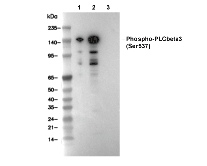 Lane 1: 293T, Lane 2: 293T (Anisomycin, 25 ug/ml, 30 min), Lane 3: 293T (Anisomycin, 25 ug/ml, 15 min; λ phosphatase treated)
Experiment Essentials
WB
Recommended SDS-PAGE separating gel concentration: 5%.
Exposure time of at least 60s is recommended.
Recommended SDS-PAGE separating gel concentration: 5%.
Exposure time of at least 60s is recommended.
Usage Information
| Dilution |
|---|
|
| Application |
|---|
| WB |
| Reactivity |
|---|
| Human, Mouse, Rat |
| Source |
|---|
| Rabbit Monoclonal Antibody |
| Storage Buffer |
|---|
| PBS, pH 7.2+50% Glycerol+0.05% BSA+0.01% NaN3 |
| Storage (from the date of receipt) |
|---|
| -20°C (avoid freeze-thaw cycles), 2 years |
| Predicted MW |
|---|
| 150 kDa |
| Positive Control | HeLa cells (Anisomycin, 20 μg/ml, 15 min) |
|---|---|
| Negative Control |
Experimental Methods
| WB |
|---|
Experimental Protocol:
Sample preparation
1. Tissue: Lyse the tissue sample by adding an appropriate volume of ice-cold RIPA/NP-40 Lysis Buffer (containing Protease Inhibitor Cocktail, Phosphatase Inhibitor Cocktail),and homogenize the tissue at a low temperature or lyse it by sonication on ice, then incubate on ice for 30 minutes. 2. Adherent cell: Aspirate the culture medium and wash the cells with ice-cold PBS twice. Lyse the cells by adding an appropriate volume of RIPA/NP-40 Lysis Buffer (containing Protease Inhibitor Cocktail, Phosphatase Inhibitor Cocktail) , sonicate to lyse the cells, and incubate on ice for 30 minutes. 3. Suspension cell: Transfer the culture medium to a pre-cooled centrifuge tube. Centrifuge and aspirate the supernatant. Wash the cells with ice-cold PBS twice. Lyse the cells by adding an appropriate volume of RIPA/NP-40 Lysis Buffer (containing Protease Inhibitor Cocktail, Phosphatase Inhibitor Cocktail) , sonicate to lyse the cells, and incubate on ice for 30 minutes. 4. Place the lysate into a pre-cooled microcentrifuge tube. Centrifuge at 4°C for 15 min. Collect the supernatant;
5. Remove a small volume of lysate to determine the protein concentration;
6. Combine the lysate with protein loading buffer. Boil 20 µL sample under 95-100°C for 5 min. Centrifuge for 5 min after cool down on ice.
Electrophoretic separation
1. According to the concentration of extracted protein, load appropriate amount of protein sample and marker onto SDS-PAGE gels for electrophoresis. Recommended separating gel (lower gel) concentration: 5%. Reference Table for Selecting SDS-PAGE Separation Gel Concentrations 2. Power up 80V for 30 minutes. Then the power supply is adjusted (110 V~150 V), the Marker is observed, and the electrophoresis can be stopped when the indicator band of the predyed protein Marker where the protein is located is properly separated. (Note that the current should not be too large when electrophoresis, too large current (more than 150 mA) will cause the temperature to rise, affecting the result of running glue. If high currents cannot be avoided, an ice bath can be used to cool the bath.)
Transfer membrane
1. Take out the converter, soak the clip and consumables in the pre-cooled converter;
2. Activate PVDF membrane with methanol for 1 min and rinse with transfer buffer;
3. Install it in the order of "black edge of clip - sponge - filter paper - filter paper - glue -PVDF membrane - filter paper - filter paper - sponge - white edge of clip"; 4. The protein was electrotransferred to PVDF membrane. ( 0.45 µm PVDF membrane is recommended ) Reference Table for Selecting PVDF Membrane Pore Size Specifications Recommended conditions for wet transfer: 200 mA, 120 min. ( Note that the transfer conditions can be adjusted according to the protein size. For high-molecular-weight proteins, a higher current and longer transfer time are recommended. However, ensure that the transfer tank remains at a low temperature to prevent gel melting.)
Block
1. After electrotransfer, wash the film with TBST at room temperature for 5 minutes;
2. Incubate the film in the blocking solution ( recommending 5% BSA solution)
for 1 hour at room temperature;
3. Wash the film with TBST for 3 times, 5 minutes each time.
Antibody incubation
1. Use primary antibody dilution buffer to prepare the primary antibody working liquid (recommended dilution ratio for primary antibody 1:1000), gently shake and incubate with the film at 4°C overnight; 2. Wash the film with TBST 3 times, 5 minutes each time;
3. Add the secondary antibody to the blocking solution and incubate with the film gently at room temperature for 1 hour;
4. After incubation, wash the film with TBST 3 times for 5 minutes each time.
Antibody staining
1. Add the prepared ECL luminescent substrate (or select other color developing substrate according to the second antibody) and mix evenly;
2. Incubate with the film for 1 minute, remove excess substrate (keep the film moist), wrap with plastic film, and expose in the imaging system. (Exposure time of at least 60s is recommended) |
Biological Description
| Specificity |
|---|
| Phospho-PLCβ3 (Ser537) Antibody (Rabbit mAb) [K10E11] detects endogenous levels of total PLCβ3 protein only when it is phosphorylated at Ser537. |
| Subcellular Location |
|---|
| Cytoplasm, Membrane, Nucleus |
| Uniprot ID |
|---|
| Q01970 |
| Clone |
|---|
| K10E11 |
| Synonym(s) |
|---|
| 1-phosphatidylinositol 4,5-bisphosphate phosphodiesterase beta-3; phosphoinositide phospholipase C; Phosphoinositide phospholipase C-beta-3; phospholipase C beta 3; Phospholipase C-beta-3; phospholipase C, PLC beta 3; PLC-beta-3; PLCB3; SMDCD |
| Background |
|---|
| Phospho-PLCβ3 (Ser537) represents a functionally significant regulatory state of phospholipase C beta 3 within the phosphoinositide-specific PLC family that links G protein–coupled and receptor tyrosine kinase signaling to phosphatidylinositol 4,5-bisphosphate hydrolysis and downstream calcium–PKC pathways, with PLCβ3 catalyzing generation of inositol 1,4,5-trisphosphate and diacylglycerol in response to diverse extracellular cues, including VEGF, thrombin, and other GPCR agonists. PLCβ3 possesses an N-terminal pleckstrin homology domain followed by an EF-hand region, a tightly associated X–Y catalytic core separated by a short linker, and a C-terminal C2 domain that together coordinate membrane association, calcium binding, and substrate engagement, while C-terminal regulatory segments integrate inputs from Gαq/11, Gβγ, and protein kinases. Ser537 lies within the N-terminal half of PLCβ3 upstream of the X domain and becomes rapidly phosphorylated in endothelial cells upon VEGF stimulation through VEGFR2, with phosphorylation detectable within thirty seconds and reaching maximal levels by one minute, in parallel with phosphorylation at the inhibitory Ser1105 site and tyrosine phosphorylation of PLCγ1 at Tyr783. VEGFR2 activation by VEGF engages both classical PLCγ1 and PLCβ3 axes, and VEGFR2-selective kinase inhibition abolishes VEGF-induced phosphorylation of PLCβ3 at Ser537 and Ser1105, indicating that Ser537 modification lies downstream of the VEGF–VEGFR2 receptor tyrosine kinase cascade rather than generic GPCR signaling. PLCβ3 Ser537 phosphorylation accompanies increased catalytic PIP2 hydrolysis manifested as elevated IP1/IP3 production in VEGF-stimulated endothelial cells, and PLCβ3 knockdown reduces VEGF-induced inositol phosphate accumulation demonstrating a direct contribution of PLCβ3 to VEGF-driven phosphoinositide turnover alongside PLCγ1. VEGF–VEGFR2–PLCβ3 signaling regulates endothelial directional migration by coupling PIP2 hydrolysis and diacylglycerol production to activation of the small GTPase CDC42, with PLCβ3 depletion selectively impairing CDC42 activation while leaving RhoA and Rac1 responses intact, correlating with loss of directional migration, reduced stress fiber formation, and diminished filopodia-associated guidance structures at sprout tips during angiogenic outgrowth. VEGF stimulation induces PLCβ3 recruitment and phosphorylation in sprouting endothelial cells in embryoid body angiogenesis models, with phosphorylated PLCβ3 localizing to CD31-positive vascular structures, indicating a role for phospho-PLCβ3 pools including the Ser537-phosphorylated fraction in angiogenic front signaling and vascular sprouting. PLCβ3 behaves as a negative regulator of endothelial proliferation, since PLCβ3 knockdown accelerates cell cycle progression with increased S-phase fraction and elevated cyclin D1 and CDC2 expression without altering VEGF-induced ERK, JNK, or p38 activation, placing Ser537-modified PLCβ3 within a signaling branch that balances proliferation against migration during angiogenesis. Both Ser537 and Ser1105 residues on PLCβ3 become phosphorylated in response to VEGF and to GPCR agonists such as thrombin and PAR ligands, situating Ser537 within a broader phosphorylation network in which Ser1105 phosphorylation by PKA or PKC suppresses PLCβ3 enzymatic activity while Ser537 phosphorylation by CaMKII contributes to basal or stimulus-tuned activity, giving phospho-PLCβ3 (Ser537) value as a readout of Ca2+/CaMKII-linked modulation of PLCβ3 within growth factor and GPCR pathways. The VEGF–VEGFR2–PLCβ3 axis cross-talks with Gq/11 and Gβγ subunits, since VEGFR2 activation engages G-protein signaling upstream of PLCβ3 in endothelium, and PLCβ3 integrates RTK and GPCR inputs at the level of phosphoinositide hydrolysis and CDC42-dependent cytoskeletal remodeling. |
| References |
|---|
|
Tech Support
Tel: +1-832-582-8158 Ext:3
If you have any other enquiries, please leave a message.

Products are for research use only. Not for human use. We do not sell to patients.
©Copyright 2013 Selleck Chemicals. All Rights Reserved.