-
 Australia
-
 Austria
-
 Belgium
-
 Brazil
-
 Canada
-
 China
China
-
 Czech Republic
-
 Denmark
-
 Finland
-
 France
-
 Germany
-
 Greece
-
 Hong Kong
-
 Hungary
-
 Iceland
-
 India
India
-
 Ireland
-
 Israel
-
 Italy
Italy
-
 Japan
Japan
-
 Korea
Korea
-
 Luxembourg
-
 Malaysia
-
 Netherlands
-
 New Zealand
-
 Norway
-
 Poland
-
 Qatar
Qatar
-
 Romania
-
 Saudi Arabia
-
 Singapore
-
 Spain
Spain
-
 Sweden
-
 Switzerland
-
 Taiwan
-
 Turkey
-
 United Kingdom
-
 United States
research use only
Phospho-MLKL (Ser345) Antibody [G22F14]
Cat.No.: F1199
Application:
Reactivity:
-
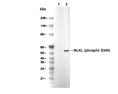 Lane 1: L-929, Lane 2: L-929 (TNFα, 20 ng/ml; BV6, 100 nM; z-VAD, 20 µM, 8 h)
Usage Information
| Dilution |
|---|
|
| Application |
|---|
| WB, IP |
| Reactivity |
|---|
| Mouse |
| Source |
|---|
| Rabbit Monoclonal Antibody |
| Storage Buffer |
|---|
| PBS, pH 7.2+50% Glycerol+0.05% BSA+0.01% NaN3 |
| Storage (from the date of receipt) |
|---|
| -20°C (avoid freeze-thaw cycles), 2 years |
| Predicted MW Observed MW |
|---|
| 54 kDa, 57 kDa 54 kDa, 53 kDa |
| *Why do the predicted and actual molecular weights differ? The following reasons may explain differences between the predicted and actual protein molecular weight. Post-translational modifications(e.g., phosphorylation, glycosylation); Splice variants and isoforms; Relative charge; Multimerization. |
| Positive Control | L-929 cells (TNF α, 20 ng/ml; Smac mimetic, 100 nM; z-VAD, 20 µM, 8 h) |
|---|---|
| Negative Control | Mouse brain tissue; Mouse colon tissue; Mouse lung tissue; Mouse retina tissue; Mouse liver tissue; L-929 cells; Raw264.7 cells |
Experimental Methods
| WB |
|---|
Experimental Protocol:
Sample preparation
1. Tissue: Lyse the tissue sample by adding an appropriate volume of ice-cold RIPA/NP-40 Lysis Buffer (containing Protease Inhibitor Cocktail, Phosphatase Inhibitor Cocktail),and homogenize the tissue at a low temperature. 2. Adherent cell: Aspirate the culture medium and wash the cells with ice-cold PBS twice. Lyse the cells by adding an appropriate volume of RIPA/NP-40 Lysis Buffer (containing Protease Inhibitor Cocktail, Phosphatase Inhibitor Cocktail) and put the sample on ice for 5 min. 3. Suspension cell: Transfer the culture medium to a pre-cooled centrifuge tube. Centrifuge and aspirate the supernatant. Wash the cells with ice-cold PBS twice. Lyse the cells by adding an appropriate volume of RIPA/NP-40 Lysis Buffer (containing Protease Inhibitor Cocktail, Phosphatase Inhibitor Cocktail) and put the sample on ice for 5 min. 4. Place the lysate into a pre-cooled microcentrifuge tube. Centrifuge at 4°C for 15 min. Collect the supernatant;
5. Remove a small volume of lysate to determine the protein concentration;
6. Combine the lysate with protein loading buffer. Boil 20 µL sample under 95-100°C for 5 min. Centrifuge for 5 min after cool down on ice.
Electrophoretic separation
1. According to the concentration of extracted protein, load appropriate amount of protein sample and marker onto SDS-PAGE gels for electrophoresis. Recommended separating gel (lower gel) concentration: 10%. Reference Table for Selecting SDS-PAGE Separation Gel Concentrations 2. Power up 80V for 30 minutes. Then the power supply is adjusted (110 V~150 V), the Marker is observed, and the electrophoresis can be stopped when the indicator band of the predyed protein Marker where the protein is located is properly separated. (Note that the current should not be too large when electrophoresis, too large current (more than 150 mA) will cause the temperature to rise, affecting the result of running glue. If high currents cannot be avoided, an ice bath can be used to cool the bath.)
Transfer membrane
1. Take out the converter, soak the clip and consumables in the pre-cooled converter;
2. Activate PVDF membrane with methanol for 1 min and rinse with transfer buffer;
3. Install it in the order of "black edge of clip - sponge - filter paper - filter paper - glue -PVDF membrane - filter paper - filter paper - sponge - white edge of clip"; 4. The protein was electrotransferred to PVDF membrane. ( 0.45 µm PVDF membrane is recommended ) Reference Table for Selecting PVDF Membrane Pore Size Specifications Recommended conditions for wet transfer: 200 mA, 120 min. ( Note that the transfer conditions can be adjusted according to the protein size. For high-molecular-weight proteins, a higher current and longer transfer time are recommended. However, ensure that the transfer tank remains at a low temperature to prevent gel melting.)
Block
1. After electrotransfer, wash the film with TBST at room temperature for 5 minutes;
2. Incubate the film in the blocking solution ( recommending 5% BSA solution)
for 1 hour at room temperature;
3. Wash the film with TBST for 3 times, 5 minutes each time.
Antibody incubation
1. Use 5% skim milk powder to prepare the primary antibody working liquid (recommended dilution ratio for primary antibody 1:1000), gently shake and incubate with the film at 4°C overnight; 2. Wash the film with TBST 3 times, 5 minutes each time;
3. Add the secondary antibody to the blocking solution and incubate with the film gently at room temperature for 1 hour;
4. After incubation, wash the film with TBST 3 times for 5 minutes each time.
Antibody staining
1. Add the prepared ECL luminescent substrate (or select other color developing substrate according to the second antibody) and mix evenly;
2. Incubate with the film for 1 minute, remove excess substrate (keep the film moist), wrap with plastic film, and expose in the imaging system. |
Biological Description
| Specificity |
|---|
| Phospho-MLKL (Ser345) Antibody [G22F14] detects endogenous levels of total MLKL protein only when it is phosphorylated at Ser345. |
| Subcellular Location |
|---|
| Cell membrane, Cytoplasm, Membrane, Nucleus |
| Uniprot ID |
|---|
| Q8NB16 |
| Clone |
|---|
| G22F14 |
| Synonym(s) |
|---|
| Mixed lineage kinase domain-like protein; Mlkl |
| Background |
|---|
| Phospho-MLKL (Ser345) is a pseudokinase within the mixed lineage kinase domain-like protein family, serving as a central effector of necroptosis, a regulated form of necrotic cell death activated by TNF family cytokines, toll-like receptors, and viral sensors when caspase activity is blocked. Mouse MLKL is a 464-amino-acid protein with an N-terminal four-helix bundle (4HB; residues 1–140), a flexible linker, and a C-terminal pseudokinase domain (PSKD; residues 175–425) that lacks catalytic activity due to a non-canonical Asp381. The critical Ser345 residue resides in the activation loop of the PSKD. Necrosome assembly, mediated by RIPK1/RIPK3 interactions through RHIM domains, enables RIPK3 to phosphorylate MLKL at Ser345. This phosphorylation induces a conformational change that disrupts Lys219-Gln343 interactions, exposing the 4HB for homotrimerization. Phospho-MLKL trimers then translocate from the cytosol to the plasma membrane, binding phosphatidylinositol phosphates (like PI(4,5)P2 and InsP6) through basic pockets in the 4HB, and form membrane pores that cause calcium influx and osmotic lysis of the cell. During viral infection, nuclear ZBP1 recruits RIPK3 to phosphorylate MLKL at Ser345, leading to nuclear envelope rupture and DNA leakage, which activates cytosolic antiviral responses. The coiled-coil domain 2 (CC2; residues 120–140) promotes MLKL oligomerization, while PGAM5 anchors the necrosome to mitochondria for efficient execution. Ser345 phosphorylation is indispensable, mutation at this site abolishes necroptosis but not apoptosis, making phospho-MLKL a biomarker of necroptosis detectable in tissue via immunohistochemistry, especially in Casp8/FADD-deficient models showing keratinocyte or intestinal necroptosis. |
| References |
|---|
|
Tech Support
Tel: +1-832-582-8158 Ext:3
If you have any other enquiries, please leave a message.
Products are for research use only. Not for human use. We do not sell to patients.
©Copyright 2013 Selleck Chemicals. All Rights Reserved.