
-
 Australia
-
 Austria
-
 Belgium
-
 Brazil
-
 Canada
-
 China
China
-
 Czech Republic
-
 Denmark
-
 Finland
-
 France
-
 Germany
-
 Greece
-
 Hong Kong
-
 Hungary
-
 Iceland
-
 India
India
-
 Ireland
-
 Israel
-
 Italy
Italy
-
 Japan
Japan
-
 Korea
Korea
-
 Luxembourg
-
 Malaysia
-
 Netherlands
-
 New Zealand
-
 Norway
-
 Poland
-
 Qatar
Qatar
-
 Romania
-
 Saudi Arabia
-
 Singapore
-
 Spain
Spain
-
 Sweden
-
 Switzerland
-
 Taiwan
-
 Turkey
-
 United Kingdom
-
 United States
research use only
Phospho-FoxM1 (Thr600) Antibody (Rabbit mAb) [A10D13]
CatNo: F1412
Application:
Reactivity:
-
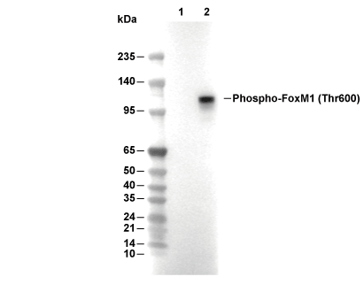 Lane 1: Hela, Lane 2: Hela (Nocodazole, 100 ng/ml, 24 h)
Usage Information
| Dilution |
|---|
|
| Application |
|---|
| WB, IP |
| Reactivity |
|---|
| Human |
| Source |
|---|
| Rabbit Monoclonal Antibody |
| Storage Buffer |
|---|
| PBS, pH 7.2+50% Glycerol+0.05% BSA+0.01% NaN3 |
| Storage (from the date of receipt) |
|---|
| -20°C (avoid freeze-thaw cycles), 2 years |
| Predicted MW |
|---|
| 84 kDa |
| Positive Control | HeLa cells (synchronized in mitosis with Nocodazole, 100 ng/ml, 16 h) |
|---|---|
| Negative Control | HeLa cells |
Experimental Methods
| WB |
|---|
Experimental Protocol:
Sample preparation
1. Tissue: Lyse the tissue sample by adding an appropriate volume of ice-cold RIPA/NP-40 Lysis Buffer (containing Protease Inhibitor Cocktail, Phosphatase Inhibitor Cocktail),and homogenize the tissue at a low temperature or lyse it by sonication on ice, then incubate on ice for 30 minutes. 2. Adherent cell: Aspirate the culture medium and wash the cells with ice-cold PBS twice. Lyse the cells by adding an appropriate volume of RIPA/NP-40 Lysis Buffer (containing Protease Inhibitor Cocktail, Phosphatase Inhibitor Cocktail) , sonicate to lyse the cells, and incubate on ice for 30 minutes. 3. Suspension cell: Transfer the culture medium to a pre-cooled centrifuge tube. Centrifuge and aspirate the supernatant. Wash the cells with ice-cold PBS twice. Lyse the cells by adding an appropriate volume of RIPA/NP-40 Lysis Buffer (containing Protease Inhibitor Cocktail, Phosphatase Inhibitor Cocktail) , sonicate to lyse the cells, and incubate on ice for 30 minutes. 4. Place the lysate into a pre-cooled microcentrifuge tube. Centrifuge at 4°C for 15 min. Collect the supernatant;
5. Remove a small volume of lysate to determine the protein concentration;
6. Combine the lysate with protein loading buffer. Boil 20 µL sample under 95-100°C for 5 min. Centrifuge for 5 min after cool down on ice.
Electrophoretic separation
1. According to the concentration of extracted protein, load appropriate amount of protein sample and marker onto SDS-PAGE gels for electrophoresis. Recommended separating gel (lower gel) concentration: 10%. Reference Table for Selecting SDS-PAGE Separation Gel Concentrations 2. Power up 80V for 30 minutes. Then the power supply is adjusted (110 V~150 V), the Marker is observed, and the electrophoresis can be stopped when the indicator band of the predyed protein Marker where the protein is located is properly separated. (Note that the current should not be too large when electrophoresis, too large current (more than 150 mA) will cause the temperature to rise, affecting the result of running glue. If high currents cannot be avoided, an ice bath can be used to cool the bath.)
Transfer membrane
1. Take out the converter, soak the clip and consumables in the pre-cooled converter;
2. Activate PVDF membrane with methanol for 1 min and rinse with transfer buffer;
3. Install it in the order of "black edge of clip - sponge - filter paper - filter paper - glue -PVDF membrane - filter paper - filter paper - sponge - white edge of clip"; 4. The protein was electrotransferred to PVDF membrane. ( 0.45 µm PVDF membrane is recommended ) Reference Table for Selecting PVDF Membrane Pore Size Specifications Recommended conditions for wet transfer: 200 mA, 120 min. ( Note that the transfer conditions can be adjusted according to the protein size. For high-molecular-weight proteins, a higher current and longer transfer time are recommended. However, ensure that the transfer tank remains at a low temperature to prevent gel melting.)
Block
1. After electrotransfer, wash the film with TBST at room temperature for 5 minutes;
2. Incubate the film in the blocking solution ( recommending 5% BSA solution)
for 1 hour at room temperature;
3. Wash the film with TBST for 3 times, 5 minutes each time.
Antibody incubation
1. Use 5% skim milk powder to prepare the primary antibody working liquid (recommended dilution ratio for primary antibody 1:1000), gently shake and incubate with the film at 4°C overnight; 2. Wash the film with TBST 3 times, 5 minutes each time;
3. Add the secondary antibody to the blocking solution and incubate with the film gently at room temperature for 1 hour;
4. After incubation, wash the film with TBST 3 times for 5 minutes each time.
Antibody staining
1. Add the prepared ECL luminescent substrate (or select other color developing substrate according to the second antibody) and mix evenly;
2. Incubate with the film for 1 minute, remove excess substrate (keep the film moist), wrap with plastic film, and expose in the imaging system. |
Biological Description
| Specificity |
|---|
| Phospho-FoxM1 (Thr600) Antibody (Rabbit mAb) [A10D13] detects endogenous levels of FoxM1 protein only when phosphorylated at Thr600. |
| Subcellular Location |
|---|
| Cytoplasm, Nucleus |
| Uniprot ID |
|---|
| Q08050 |
| Clone |
|---|
| A10D13 |
| Synonym(s) |
|---|
| FKHL16; forkhead box M1; forkhead box M1-D; Forkhead box protein M1; FOXM1; FOXM1A; FOXM1B; FOXM1C; HFH-11; HFH11; HNF-3; MPP2; PIG29; TGT3; Transcription factor Trident; TRIDENT; WIN |
| Background |
|---|
| Phosphorylated FoxM1 at Thr600 represents a mitosis‑linked activated form of the Forkhead box M1 transcription factor, a member of the forkhead family that controls expression of a wide panel of cell cycle genes essential for DNA replication, G2/M transition and orderly mitotic progression. FoxM1 contains an N‑terminal transactivation and regulatory region with multiple serine/threonine phosphorylation sites, a centrally located forkhead DNA‑binding domain that recognizes consensus sequences in promoters of cell cycle genes, and a C‑terminal transactivation domain that recruits co‑activators; coordinated phosphorylation by Cyclin/Cdk complexes at sites including Thr600, Thr611 and Thr620, together with other kinase inputs, switches FoxM1 from an inactive to a transcriptionally competent state during late S and G2/M. Thr600 lies within a Cdk consensus motif in the transactivation region, and mutational and interaction analyses show that phosphorylation of Thr600 and Thr611 enhances FoxM1 binding to the cell cycle phosphatase CDC25A, stabilizes this interaction and promotes CDC25A‑dependent dephosphorylation of inhibitory Cdk residues, thereby closing a feed‑forward loop in which FoxM1 activation and Thr600 phosphorylation reinforce Cdk1/Cyclin B activation and drive entry into mitosis. This phosphorylation state is cell cycle–regulated, with FoxM1 protein levels rising from G1/S and Thr600‑phosphorylated FoxM1 accumulating in late S/G2, just before nuclear translocation and peak transcriptional activity on promoters such as CCNB1, PLK1, CDC25B, Aurora B and survivin, which collectively control centrosome maturation, spindle formation, chromosome segregation and cytokinesis. Phospho‑specific antibodies and HTRF detection kits that recognize FoxM1 only when phosphorylated at Thr600 demonstrate that Thr600 phosphorylation is tightly coupled to mitotic entry and is increased in proliferating and tumor cells, and these tools are widely employed as biomarkers of FoxM1 pathway activation and for screening small‑molecule FoxM1 inhibitors that suppress Thr600 phosphorylation and downregulate FoxM1 target genes. Reviews of FoxM1 biology in cancer show that FOXM1 is overexpressed and hyperactivated in many human tumors, including breast, lung, liver, pancreas and prostate cancers, and contributes to multiple hallmarks of malignancy such as uncontrolled proliferation, genome instability, epithelial–mesenchymal transition, metastasis, therapy resistance and evasion of antitumor immunity, in part by sustaining expression of G2/M regulators, DNA repair factors and stemness‑associated genes under the control of phosphorylated FoxM1. High levels of Thr600‑phosphorylated FoxM1 correlate with aggressive clinicopathologic features and poor prognosis, and experimental FoxM1 inhibitors that reduce Thr600 phosphorylation and interfere with FoxM1–CDC25A and FoxM1–PLK1 circuits show suppression of tumor xenograft growth and decreased expression of FoxM1‑regulated mitotic genes, supporting the use of phospho‑FoxM1 Thr600 both as a mechanistic readout of on‑target drug action and as a candidate therapeutic biomarker. |
| References |
|---|
|
Tech Support
Tel: +1-832-582-8158 Ext:3
If you have any other enquiries, please leave a message.

Products are for research use only. Not for human use. We do not sell to patients.
©Copyright 2013 Selleck Chemicals. All Rights Reserved.