-
 Australia
-
 Austria
-
 Belgium
-
 Brazil
-
 Canada
-
 China
China
-
 Czech Republic
-
 Denmark
-
 Finland
-
 France
-
 Germany
-
 Greece
-
 Hong Kong
-
 Hungary
-
 Iceland
-
 India
India
-
 Ireland
-
 Israel
-
 Italy
Italy
-
 Japan
Japan
-
 Korea
Korea
-
 Luxembourg
-
 Malaysia
-
 Netherlands
-
 New Zealand
-
 Norway
-
 Poland
-
 Qatar
Qatar
-
 Romania
-
 Saudi Arabia
-
 Singapore
-
 Spain
Spain
-
 Sweden
-
 Switzerland
-
 Taiwan
-
 Turkey
-
 United Kingdom
-
 United States
research use only
Phospho-BRAF (Ser729) Antibody [H9E15]
Cat.No.: F2240
Application:
Reactivity:
-
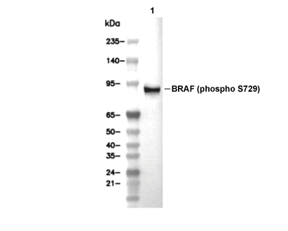 Lane 1: PC-12, Lane 2: PC-12 (λ phosphatase-treated)
Experiment Essentials
WB
Exposure time of at least 120s is recommended.
Exposure time of at least 120s is recommended.
Usage Information
| Dilution |
|---|
|
| Application |
|---|
| WB |
| Reactivity |
|---|
| Rat |
| Source |
|---|
| Rabbit Monoclonal Antibody |
| Storage Buffer |
|---|
| PBS, pH 7.2+50% Glycerol+0.05% BSA+0.01% NaN3 |
| Storage (from the date of receipt) |
|---|
| -20°C (avoid freeze-thaw cycles), 2 years |
| Predicted MW |
|---|
| 84 kDa |
| Positive Control | PC-12 cells |
|---|---|
| Negative Control |
Experimental Methods
| WB |
|---|
Experimental Protocol:
Sample preparation
1. Tissue: Lyse the tissue sample by adding an appropriate volume of ice-cold RIPA/NP-40 Lysis Buffer (containing Protease Inhibitor Cocktail, Phosphatase Inhibitor Cocktail),and homogenize the tissue at a low temperature. 2. Adherent cell: Aspirate the culture medium and wash the cells with ice-cold PBS twice. Lyse the cells by adding an appropriate volume of RIPA/NP-40 Lysis Buffer (containing Protease Inhibitor Cocktail, Phosphatase Inhibitor Cocktail) and put the sample on ice for 5 min. 3. Suspension cell: Transfer the culture medium to a pre-cooled centrifuge tube. Centrifuge and aspirate the supernatant. Wash the cells with ice-cold PBS twice. Lyse the cells by adding an appropriate volume of RIPA/NP-40 Lysis Buffer (containing Protease Inhibitor Cocktail, Phosphatase Inhibitor Cocktail) and put the sample on ice for 5 min. 4. Place the lysate into a pre-cooled microcentrifuge tube. Centrifuge at 4°C for 15 min. Collect the supernatant;
5. Remove a small volume of lysate to determine the protein concentration;
6. Combine the lysate with protein loading buffer. Boil 20 µL sample under 95-100°C for 5 min. Centrifuge for 5 min after cool down on ice.
Electrophoretic separation
1. According to the concentration of extracted protein, load appropriate amount of protein sample and marker onto SDS-PAGE gels for electrophoresis. Recommended separating gel (lower gel) concentration: 10%. Reference Table for Selecting SDS-PAGE Separation Gel Concentrations 2. Power up 80V for 30 minutes. Then the power supply is adjusted (110 V~150 V), the Marker is observed, and the electrophoresis can be stopped when the indicator band of the predyed protein Marker where the protein is located is properly separated. (Note that the current should not be too large when electrophoresis, too large current (more than 150 mA) will cause the temperature to rise, affecting the result of running glue. If high currents cannot be avoided, an ice bath can be used to cool the bath.)
Transfer membrane
1. Take out the converter, soak the clip and consumables in the pre-cooled converter;
2. Activate PVDF membrane with methanol for 1 min and rinse with transfer buffer;
3. Install it in the order of "black edge of clip - sponge - filter paper - filter paper - glue -PVDF membrane - filter paper - filter paper - sponge - white edge of clip"; 4. The protein was electrotransferred to PVDF membrane. ( 0.45 µm PVDF membrane is recommended ) Reference Table for Selecting PVDF Membrane Pore Size Specifications Recommended conditions for wet transfer: 200 mA, 120 min. ( Note that the transfer conditions can be adjusted according to the protein size. For high-molecular-weight proteins, a higher current and longer transfer time are recommended. However, ensure that the transfer tank remains at a low temperature to prevent gel melting.)
Block
1. After electrotransfer, wash the film with TBST at room temperature for 5 minutes;
2. Incubate the film in the blocking solution ( recommending 5% BSA solution)
for 1 hour at room temperature;
3. Wash the film with TBST for 3 times, 5 minutes each time.
Antibody incubation
1. Use 5% skim milk powder to prepare the primary antibody working liquid (recommended dilution ratio for primary antibody 1:1000), gently shake and incubate with the film at 4°C overnight; 2. Wash the film with TBST 3 times, 5 minutes each time;
3. Add the secondary antibody to the blocking solution and incubate with the film gently at room temperature for 1 hour;
4. After incubation, wash the film with TBST 3 times for 5 minutes each time.
Antibody staining
1. Add the prepared ECL luminescent substrate (or select other color developing substrate according to the second antibody) and mix evenly;
2. Incubate with the film for 1 minute, remove excess substrate (keep the film moist), wrap with plastic film, and expose in the imaging system. (Exposure time of at least 120s is recommended) |
Biological Description
| Specificity |
|---|
| Phospho-BRAF (Ser729) Antibody [H9E15] detects endogenous levels of total BRAF protein only when it is phosphorylated at Ser729. |
| Subcellular Location |
|---|
| Cell membrane, Membrane, Cytoplasm, Nucleus |
| Uniprot ID |
|---|
| P15056 |
| Clone |
|---|
| H9E15 |
| Synonym(s) |
|---|
| BRAF1; RAFB1; BRAF; Serine/threonine-protein kinase B-raf; Proto-oncogene B-Raf; p94; v-Raf murine sarcoma viral oncogene homolog B1 |
| Background |
|---|
| Phospho-BRAF (Ser729) belongs to the RAF family of serine/threonine kinases that relay signals from RAS to MEK in the MAPK/ERK cascade, driving cell proliferation, differentiation, and survival. BRAF comprises 766 amino acids organized into RAS-binding domain (residues 150–224), cysteine-rich domain (225–303), and kinase domain (446–723), with the C-terminal Ser729 positioned beyond the kinase domain for 14-3-3 interaction. Phosphorylation at Ser729 occurs through AMPK activation under energy stress, where AMPKa directly targets this residue within its optimal consensus motif. This modification enhances BRAF binding to 14-3-3 proteins, sequestering the kinase and stabilizing autoinhibited states by bridging pSer729 sites between BRAF protomers via a single 14-3-3 dimer. In autoinhibited complexes, 14-3-3 locks the C-terminal tails, preventing premature dimerization while preserving assembly competence for activation. Upon RAS-GTP engagement, release from this inhibitory conformation allows BRAF dimerization, with αC-helix shifting to align regulatory spines for MEK phosphorylation. Phospho-Ser729 thus toggles BRAF between autoinhibited monomers and active dimers, fine-tuning pathway output; mutation to Ala boosts KSR1 association, elevates MEK-ERK signaling, and accelerates proliferation in keratinocytes. AMPK-mediated pSer729 disrupts BRAF-KSR1 scaffolds essential for efficient signal propagation, attenuating ERK activation during AICAR-induced stress. BRAF V600E mutations in melanoma bypass such regulation, causing constitutive kinase activity and therapy resistance, while wild-type pSer729 curbs pathological hyperactivation. Released 14-3-3 in active dimers modulates MEK affinity through steric hindrance, promoting sequential substrate release. |
| References |
|---|
|
Tech Support
Tel: +1-832-582-8158 Ext:3
If you have any other enquiries, please leave a message.
Products are for research use only. Not for human use. We do not sell to patients.
©Copyright 2013 Selleck Chemicals. All Rights Reserved.