
-
 Australia
-
 Austria
-
 Belgium
-
 Brazil
-
 Canada
-
 China
China
-
 Czech Republic
-
 Denmark
-
 Finland
-
 France
-
 Germany
-
 Greece
-
 Hong Kong
-
 Hungary
-
 Iceland
-
 India
India
-
 Ireland
-
 Israel
-
 Italy
Italy
-
 Japan
Japan
-
 Korea
Korea
-
 Luxembourg
-
 Malaysia
-
 Netherlands
-
 New Zealand
-
 Norway
-
 Poland
-
 Qatar
Qatar
-
 Romania
-
 Saudi Arabia
-
 Singapore
-
 Spain
Spain
-
 Sweden
-
 Switzerland
-
 Taiwan
-
 Turkey
-
 United Kingdom
-
 United States
research use only
Phospho-BCKDHA (Ser293) Antibody (Rabbit mAb) [D4N17]
CatNo: F7105
Application:
Reactivity:
-
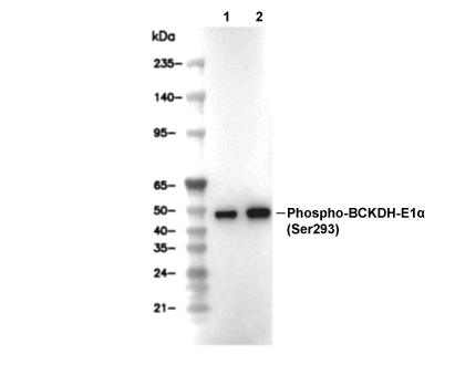 Lane 1: COS-7, Lane 2: Vero
Experiment Essentials
WB
Exposure time of at least 60s is recommended.
Exposure time of at least 60s is recommended.
Usage Information
| Dilution |
|---|
|
| Application |
|---|
| WB, IP |
| Reactivity |
|---|
| Human, Mouse, Rat, Monkey |
| Source |
|---|
| Rabbit Monoclonal Antibody |
| Storage Buffer |
|---|
| PBS, pH 7.2+50% Glycerol+0.05% BSA+0.01% NaN3 |
| Storage (from the date of receipt) |
|---|
| -20°C (avoid freeze-thaw cycles), 2 years |
| Predicted MW |
|---|
| 50 kDa |
| Positive Control | 293T cells; mIMCD-3 cells; KNRK cells; COS-7 cells; Vero cells; HeLa cells |
|---|---|
| Negative Control |
Experimental Methods
| WB |
|---|
Experimental Protocol:
Sample preparation
1. Tissue: Lyse the tissue sample by adding an appropriate volume of ice-cold RIPA/NP-40 Lysis Buffer (containing Protease Inhibitor Cocktail, Phosphatase Inhibitor Cocktail),and homogenize the tissue at a low temperature or lyse it by sonication on ice, then incubate on ice for 30 minutes. 2. Adherent cell: Aspirate the culture medium and wash the cells with ice-cold PBS twice. Lyse the cells by adding an appropriate volume of RIPA/NP-40 Lysis Buffer (containing Protease Inhibitor Cocktail, Phosphatase Inhibitor Cocktail) , sonicate to lyse the cells, and incubate on ice for 30 minutes. 3. Suspension cell: Transfer the culture medium to a pre-cooled centrifuge tube. Centrifuge and aspirate the supernatant. Wash the cells with ice-cold PBS twice. Lyse the cells by adding an appropriate volume of RIPA/NP-40 Lysis Buffer (containing Protease Inhibitor Cocktail, Phosphatase Inhibitor Cocktail) , sonicate to lyse the cells, and incubate on ice for 30 minutes. 4. Place the lysate into a pre-cooled microcentrifuge tube. Centrifuge at 4°C for 15 min. Collect the supernatant;
5. Remove a small volume of lysate to determine the protein concentration;
6. Combine the lysate with protein loading buffer. Boil 20 µL sample under 95-100°C for 5 min. Centrifuge for 5 min after cool down on ice.
Electrophoretic separation
1. According to the concentration of extracted protein, load appropriate amount of protein sample and marker onto SDS-PAGE gels for electrophoresis. Recommended separating gel (lower gel) concentration: 10%. Reference Table for Selecting SDS-PAGE Separation Gel Concentrations 2. Power up 80V for 30 minutes. Then the power supply is adjusted (110 V~150 V), the Marker is observed, and the electrophoresis can be stopped when the indicator band of the predyed protein Marker where the protein is located is properly separated. (Note that the current should not be too large when electrophoresis, too large current (more than 150 mA) will cause the temperature to rise, affecting the result of running glue. If high currents cannot be avoided, an ice bath can be used to cool the bath.)
Transfer membrane
1. Take out the converter, soak the clip and consumables in the pre-cooled converter;
2. Activate PVDF membrane with methanol for 1 min and rinse with transfer buffer;
3. Install it in the order of "black edge of clip - sponge - filter paper - filter paper - glue -PVDF membrane - filter paper - filter paper - sponge - white edge of clip"; 4. The protein was electrotransferred to PVDF membrane. ( 0.45 µm PVDF membrane is recommended ) Reference Table for Selecting PVDF Membrane Pore Size Specifications Recommended conditions for wet transfer: 200 mA, 120 min. ( Note that the transfer conditions can be adjusted according to the protein size. For high-molecular-weight proteins, a higher current and longer transfer time are recommended. However, ensure that the transfer tank remains at a low temperature to prevent gel melting.)
Block
1. After electrotransfer, wash the film with TBST at room temperature for 5 minutes;
2. Incubate the film in the blocking solution ( recommending 5% BSA solution)
for 1 hour at room temperature;
3. Wash the film with TBST for 3 times, 5 minutes each time.
Antibody incubation
1. Use primary antibody dilution buffer to prepare the primary antibody working liquid (recommended dilution ratio for primary antibody 1:1000), gently shake and incubate with the film at 4°C overnight; 2. Wash the film with TBST 3 times, 5 minutes each time;
3. Add the secondary antibody to the blocking solution and incubate with the film gently at room temperature for 1 hour;
4. After incubation, wash the film with TBST 3 times for 5 minutes each time.
Antibody staining
1. Add the prepared ECL luminescent substrate (or select other color developing substrate according to the second antibody) and mix evenly;
2. Incubate with the film for 1 minute, remove excess substrate (keep the film moist), wrap with plastic film, and expose in the imaging system. (Exposure time of at least 60s is recommended) |
Biological Description
| Specificity |
|---|
| Phospho-BCKDHA (Ser293) Antibody (Rabbit mAb) [D4N17] detects endogenous levels of BCKDHA protein only when phosphorylated at Ser292 of mature human BCKDHA, Ser293 of mature mouse BCKDHA, or Ser293 of mature rat BCKDHA. |
| Subcellular Location |
|---|
| Mitochondrion |
| Uniprot ID |
|---|
| P12694 |
| Clone |
|---|
| D4N17 |
| Synonym(s) |
|---|
| 2-oxoisovalerate dehydrogenase (lipoamide); BCKDE1A; BCKDH E1-alpha; BCKDHA; FLJ45695; MSU; MSUD1; ODBA; OVD1A |
| Background |
|---|
| Phospho-BCKDHA (Ser293) refers to the regulatory phosphorylation state of the α subunit of the branched-chain α‑keto acid dehydrogenase E1 component, a mitochondrial enzyme that catalyzes the first, rate-limiting oxidative decarboxylation step in branched-chain amino acid (BCAA) catabolism and thereby controls systemic levels of leucine, isoleucine and valine. The E1α subunit contributes the thiamine pyrophosphate–dependent decarboxylase active site and contains two serine residues that can be phosphorylated, but site 1, Ser293 in rat and equivalent residues in mouse and human, is the dominant regulatory site whose modification governs complex activity. Phosphorylation at Ser293 is carried out by BCKDH kinase (BCKDK), which recognizes residues surrounding this serine as a substrate motif; Ser293 and neighboring conserved residues such as Arg288, His292 and Asp296 are crucial for catalysis and for proper substrate recognition by BCKDK, and Ser293 substitution alters Km values for different branched-chain α‑keto acids without affecting Vmax, highlighting the dual contribution of this region to enzymatic efficiency and kinase control. When BCKDK phosphorylates E1α at Ser293, the BCKDH complex is inactivated and flux through BCAA oxidative degradation is suppressed, leading to retention of BCAAs and their α‑ketoacid derivatives; antibodies that specifically detect BCKDH-E1α only when phosphorylated at Ser293 have been used to show that high phospho-Ser293 levels correspond to low BCKDH activity in tissues. In liver, BCKDH-E1α Ser293 phosphorylation integrates nutritional and hormonal signals: hepatic BCKDH activity is markedly decreased in type 2 diabetes and obesity, and this reduction correlates with increased Ser293 phosphorylation and elevated plasma BCAA concentrations, linking phospho-E1α Ser293 to the BCAA signature associated with insulin resistance and cardiometabolic risk. Manipulation of this axis—either by inhibiting BCKDK or overexpressing the dedicated mitochondrial phosphatase PPM1K—reduces E1α Ser293 phosphorylation, reactivates BCKDH, enhances BCAA clearance and improves glucose and lipid metabolism, indicating that the Ser293 phospho-state acts as a metabolic rheostat for BCAA disposal and downstream metabolic homeostasis. Upstream signaling pathways can modulate Ser293 phosphorylation: for example, G protein–coupled oestrogen receptor (GPER) activation stimulates JNK-dependent phosphorylation of BCKDH-E1α at Ser293, and JNK silencing abolishes this GPER-induced modification, pointing to a route by which hormonal cues and stress kinases feed into BCKDH regulation via the Ser293 site. In cardiometabolic disease models, dysregulated extramitochondrial BCKDH and uncoupling of BCAA metabolism from energy production associate with altered BCKDH-E1α phosphorylation and impaired energetics, further underscoring the importance of Ser293 status for tissue-specific BCAA handling and energy balance. The thiamine-dependent decarboxylase structure of E1α, the unique regulatory role of Ser293 phosphorylation by BCKDK and dephosphorylation by PPM1K, and the documented links between phospho-BCKDH-E1α (Ser293), BCAA accumulation, diabetes, and cardiometabolic dysfunction. |
| References |
|---|
|
Tech Support
Tel: +1-832-582-8158 Ext:3
If you have any other enquiries, please leave a message.

Products are for research use only. Not for human use. We do not sell to patients.
©Copyright 2013 Selleck Chemicals. All Rights Reserved.