
-
 Australia
-
 Austria
-
 Belgium
-
 Brazil
-
 Canada
-
 China
China
-
 Czech Republic
-
 Denmark
-
 Finland
-
 France
-
 Germany
-
 Greece
-
 Hong Kong
-
 Hungary
-
 Iceland
-
 India
India
-
 Ireland
-
 Israel
-
 Italy
Italy
-
 Japan
Japan
-
 Korea
Korea
-
 Luxembourg
-
 Malaysia
-
 Netherlands
-
 New Zealand
-
 Norway
-
 Poland
-
 Qatar
Qatar
-
 Romania
-
 Saudi Arabia
-
 Singapore
-
 Spain
Spain
-
 Sweden
-
 Switzerland
-
 Taiwan
-
 Turkey
-
 United Kingdom
-
 United States
research use only
Perilipin-2 Antibody (Rabbit mAb) [K24J8]
CatNo: F5935
Application:
Reactivity:
-
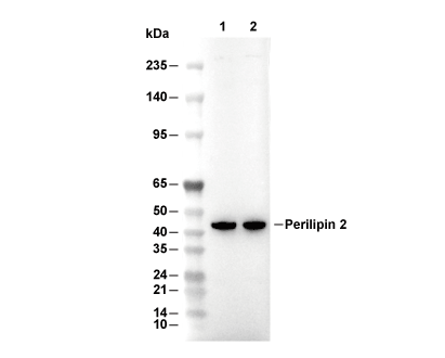 Lane 1: Mouse brain, Lane 2: Rat brain
Usage Information
| Dilution |
|---|
|
| Application |
|---|
| WB |
| Reactivity |
|---|
| Mouse, Rat, Human |
| Source |
|---|
| Rabbit Monoclonal Antibody |
| Storage Buffer |
|---|
| PBS, pH 7.2+50% Glycerol+0.05% BSA+0.01% NaN3 |
| Storage (from the date of receipt) |
|---|
| -20°C (avoid freeze-thaw cycles), 2 years |
| Predicted MW |
|---|
| 48 kDa |
| Positive Control | Human fetal liver; Rat brain; Mouse brain; Human milk; HepG2 cells; JAR cells |
|---|---|
| Negative Control |
Experimental Methods
| WB |
|---|
Experimental Protocol:
Sample preparation
1. Tissue: Lyse the tissue sample by adding an appropriate volume of ice-cold RIPA/NP-40 Lysis Buffer (containing Protease Inhibitor Cocktail),and homogenize the tissue at a low temperature or lyse it by sonication on ice, then incubate on ice for 30 minutes. 2. Adherent cell: Aspirate the culture medium and wash the cells with ice-cold PBS twice. Lyse the cells by adding an appropriate volume of RIPA/NP-40 Lysis Buffer (containing Protease Inhibitor Cocktail) , sonicate to lyse the cells, and incubate on ice for 30 minutes. 3. Suspension cell: Transfer the culture medium to a pre-cooled centrifuge tube. Centrifuge and aspirate the supernatant. Wash the cells with ice-cold PBS twice. Lyse the cells by adding an appropriate volume of RIPA/NP-40 Lysis Buffer (containing Protease Inhibitor Cocktail) , sonicate to lyse the cells, and incubate on ice for 30 minutes. 4. Place the lysate into a pre-cooled microcentrifuge tube. Centrifuge at 4°C for 15 min. Collect the supernatant;
5. Remove a small volume of lysate to determine the protein concentration;
6. Combine the lysate with protein loading buffer. Boil 20 µL sample under 95-100°C for 5 min. Centrifuge for 5 min after cool down on ice.
Electrophoretic separation
1. According to the concentration of extracted protein, load appropriate amount of protein sample and marker onto SDS-PAGE gels for electrophoresis. Recommended separating gel (lower gel) concentration: 10%. Reference Table for Selecting SDS-PAGE Separation Gel Concentrations 2. Power up 80V for 30 minutes. Then the power supply is adjusted (110 V~150 V), the Marker is observed, and the electrophoresis can be stopped when the indicator band of the predyed protein Marker where the protein is located is properly separated. (Note that the current should not be too large when electrophoresis, too large current (more than 150 mA) will cause the temperature to rise, affecting the result of running glue. If high currents cannot be avoided, an ice bath can be used to cool the bath.)
Transfer membrane
1. Take out the converter, soak the clip and consumables in the pre-cooled converter;
2. Activate PVDF membrane with methanol for 1 min and rinse with transfer buffer;
3. Install it in the order of "black edge of clip - sponge - filter paper - filter paper - glue -PVDF membrane - filter paper - filter paper - sponge - white edge of clip"; 4. The protein was electrotransferred to PVDF membrane. ( 0.45 µm PVDF membrane is recommended ) Reference Table for Selecting PVDF Membrane Pore Size Specifications Recommended conditions for wet transfer: 200 mA, 120 min. ( Note that the transfer conditions can be adjusted according to the protein size. For high-molecular-weight proteins, a higher current and longer transfer time are recommended. However, ensure that the transfer tank remains at a low temperature to prevent gel melting.)
Block
1. After electrotransfer, wash the film with TBST at room temperature for 5 minutes;
2. Incubate the film in the blocking solution for 1 hour at room temperature;
3. Wash the film with TBST for 3 times, 5 minutes each time.
Antibody incubation
1. Use primary antibody dilution buffer to prepare the primary antibody working liquid (recommended dilution ratio for primary antibody 1:1000), gently shake and incubate with the film at 4°C overnight; 2. Wash the film with TBST 3 times, 5 minutes each time;
3. Add the secondary antibody to the blocking solution and incubate with the film gently at room temperature for 1 hour;
4. After incubation, wash the film with TBST 3 times for 5 minutes each time.
Antibody staining
1. Add the prepared ECL luminescent substrate (or select other color developing substrate according to the second antibody) and mix evenly;
2. Incubate with the film for 1 minute, remove excess substrate (keep the film moist), wrap with plastic film, and expose in the imaging system. |
Biological Description
| Specificity |
|---|
| Perilipin-2 Antibody (Rabbit mAb) [K24J8] detects endogenous levels of total Perilipin-2 protein. |
| Subcellular Location |
|---|
| Lipid droplet, Membrane |
| Uniprot ID |
|---|
| Q99541 |
| Clone |
|---|
| K24J8 |
| Synonym(s) |
|---|
| ADFP, PLIN2, Perilipin-2, Adipophilin, Adipose differentiation-related protein, ADRP |
| Background |
|---|
| Perilipin‑2, also known as adipose differentiation–related protein (ADFP or adipophilin), is a member of the perilipin/PAT family of cytoplasmic lipid droplet–binding proteins and is ubiquitously expressed, with particularly high levels in tissues that handle neutral lipid flux such as liver, adipose tissue, skeletal muscle and pancreatic islets, where it acts as a structural component of lipid droplets required for their formation and maintenance. The protein contains an N‑terminal PAT domain that is highly conserved within the perilipin family and stabilizes nascent lipid droplets, a central region with 11‑mer repeats that mediate cytosolic lipid droplet binding, and a C‑terminal four‑helix bundle that supports membrane association, creating a modular architecture that allows perilipin‑2 to coat the droplet surface together with phospholipids and define the interface at which lipases and trafficking factors access stored triglycerides and cholesteryl esters. Mechanistically, perilipin‑2 promotes fatty acid uptake and storage by facilitating the sequestration of neutral lipids into cytosolic triglycerides, inhibiting their incorporation into very low‑density lipoproteins and slowing triacylglycerol turnover; genetic deletion of Adfp in mice reduces hepatic triglyceride content by 60%, increases hepatic VLDL secretion, relieves hepatosteatosis and improves whole‑body insulin resistance in leptin‑deficient animals, indicating that perilipin‑2 acts as a gatekeeper that favors intracellular lipid retention and limits export under conditions of nutrient excess. In pancreatic islets, perilipin‑2 regulates lipid handling by controlling islet triglyceride stores and influencing insulin secretion: increased perilipin‑2 expression enhances lipid accumulation and modifies the balance between nutrient signaling and lipotoxic stress, while knockdown experiments show that lowering perilipin‑2 reduces triglyceride stores and alters fatty acid‑stimulated insulin release, linking droplet scaffolding directly to β‑cell functional responses. At the level of lipolytic machinery, perilipin‑2 competes with and reduces the lipid droplet association of adipose triglyceride lipase (ATGL), thereby slowing triacylglycerol turnover and stabilizing stored fat; droplets coated predominantly with perilipin‑2 are more protective of lipids and relatively permissive to basal lipolysis compared with droplets coated by other perilipin family members, contributing to tissue‑specific differences in lipase access and energy mobilization. Peroxisome proliferator‑activated receptor‑α agonists and fasting induce perilipin‑2 expression in liver, where the protein ensures adequate lipid droplet formation to buffer incoming fatty acids and prevent uncontrolled lipotoxicity, but chronic upregulation in obesity and metabolic syndrome is associated with excessive hepatic lipid storage, nonalcoholic fatty liver disease and systemic insulin resistance. Human genetic variation in the C‑terminal region of perilipin‑2, such as a serine‑to‑proline substitution at position 251 that disrupts predicted α‑helical structure, reduces plasma triglycerides and alters lipolysis, reinforcing the importance of the helical bundle in controlling lipid droplet dynamics and triglyceride turnover. Elevated perilipin‑2 expression has also been reported in colorectal cancer, renal carcinoma and lung adenocarcinoma, where it correlates with increased lipid droplet abundance and can promote cell proliferation via Akt phosphorylation, supporting its use as a biomarker of lipid‑rich tumors and highlighting the intersection between lipid storage machinery and oncogenic signaling. |
| References |
|---|
|
Tech Support
Tel: +1-832-582-8158 Ext:3
If you have any other enquiries, please leave a message.

Products are for research use only. Not for human use. We do not sell to patients.
©Copyright 2013 Selleck Chemicals. All Rights Reserved.