-
 Australia
-
 Austria
-
 Belgium
-
 Brazil
-
 Canada
-
 China
China
-
 Czech Republic
-
 Denmark
-
 Finland
-
 France
-
 Germany
-
 Greece
-
 Hong Kong
-
 Hungary
-
 Iceland
-
 India
India
-
 Ireland
-
 Israel
-
 Italy
Italy
-
 Japan
Japan
-
 Korea
Korea
-
 Luxembourg
-
 Malaysia
-
 Netherlands
-
 New Zealand
-
 Norway
-
 Poland
-
 Qatar
Qatar
-
 Romania
-
 Saudi Arabia
-
 Singapore
-
 Spain
Spain
-
 Sweden
-
 Switzerland
-
 Taiwan
-
 Turkey
-
 United Kingdom
-
 United States
research use only
Otoferlin Antibody [J18F12]
Cat.No.: F2110
Application:
Reactivity:
-
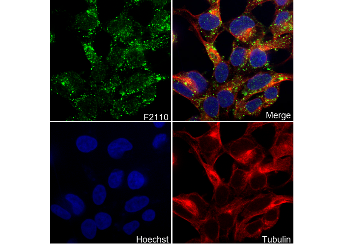 Immunofluorescent analysis of NTERA-2 cells using F2110 (green, 1:200), Hoechst (blue) and tubulin (Red).
Usage Information
| Dilution |
|---|
|
| Application |
|---|
| IF, FCM |
| Reactivity |
|---|
| Mouse, Human |
| Source |
|---|
| Mouse Monoclonal Antibody |
| Storage Buffer |
|---|
| PBS, pH 7.2+50% Glycerol+0.05% BSA+0.01% NaN3 |
| Storage (from the date of receipt) |
|---|
| -20°C (avoid freeze-thaw cycles), 2 years |
| Positive Control | Mouse inner ear tissue; SHSY-5Y cells |
|---|---|
| Negative Control |
Experimental Methods
| IF |
|---|
Experimental Protocol:
Sample Preparation
1. Adherent Cells: Place a clean, sterile coverslip in a culture dish. Once the cells grow to near confluence as a monolayer, remove the coverslip for further use.
2. Suspension Cells: Seed the cells onto a clean, sterile slide coated with poly-L-lysine.
3. Frozen Sections: Allow the slide to thaw at room temperature. Wash it with pure water or PBS for 2 times, 3 minutes each time.
4. Paraffin Sections: Deparaffinization and rehydration. Wash the slide with pure water or PBS for 3 times, 3 minutes each time. Then perform antigen retrieval.
Fixation
1. Fix the cell coverslips/spots or tissue sections at room temperature using a fixative such as 4% paraformaldehyde (4% PFA) for 10-15 minutes.
2. Wash the sample with PBS for 3 times, 3 minutes each time.
Permeabilization
1.Add a detergent such as 0.1–0.3% Triton X-100 to the sample and incubate at room temperature for 10–20 minutes.
(Note: This step is only required for intracellular antigens. For antigens expressed on the cell membrane, this step is unnecessary.)
Wash the sample with PBS for 3 times, 3 minutes each time.
Blocking
Add blocking solution and incubate at room temperature for at least 1 hour. (Common blocking solutions include: serum from the same source as the secondary antibody, BSA, or goat serum.)
Note: Ensure the sample remains moist during and after the blocking step to prevent drying, which can lead to high background.
Immunofluorescence Staining (Day 1)
1. Remove the blocking solution and add the diluted primary antibody.
2. Incubate the sample in a humidified chamber at 4°C overnight.
Immunofluorescence Staining (Day 2)
1. Remove the primary antibody and wash with PBST for 3 times, 5 minutes each time.
2. Add the diluted fluorescent secondary antibody and incubate in the dark at 4°C for 1–2 hours.
3. Remove the secondary antibody and wash with PBST for 3 times, 5 minutes each time.
4. Add diluted DAPI and incubate at room temperature in the dark for 5–10 minutes.
5. Wash with PBST for 3 times, 5 minutes each time.
Mounting
1. Mount the sample with an anti-fade mounting medium.
2. Allow the slide to dry at room temperature overnight in the dark.
3. Store the slide in a slide storage box at 4°C, protected from light.
|
Biological Description
| Specificity |
|---|
| Otoferlin Antibody [J18F12] detects endogenous levels of total Otoferlin protein. |
| Subcellular Location |
|---|
| Cell projection, Endoplasmic reticulum, Golgi apparatus, Membrane, Synapse |
| Uniprot ID |
|---|
| Q9HC10 |
| Clone |
|---|
| J18F12 |
| Synonym(s) |
|---|
| FER1L2; OTOF; Otoferlin; Fer-1-like protein 2 |
| Background |
|---|
| Otoferlin, encoded by the OTOF gene, is a large multi-C2 domain protein in the ferlin family that is primarily expressed in cochlear inner hair cells (IHCs). It acts as a key calcium sensor for synaptic vesicle exocytosis at ribbon synapses, enabling rapid sound encoding and neurotransmission. Otoferlin contains six cytosolic C2 domains (C2A-F); among these, the C2B-F domains bind Ca²⁺ through aspartate-rich loops and interact with phospholipids such as phosphatidylserine and PIP₂, as well as SNARE proteins like syntaxin-1 and SNAP-25. It also includes a FerA domain (a four-helix bundle) that provides additional Ca²⁺-dependent lipid-binding capacity and a C-terminal transmembrane domain (TMD), which is alternatively spliced (TMD1 in cochlea, TMD2 in brain) to anchor otoferlin to vesicle or plasma membranes. Through these features, otoferlin facilitates Ca²⁺-triggered glutamate vesicle fusion in concert with L-type Caᵥ1.3 channels, supports vesicle replenishment and endocytosis, and maintains high-fidelity auditory signaling across a wide range of intensities, distinguishing it from synaptotagmin in slower synapses. Mutations in OTOF, such as I318N in C2B or p.Ile515Thr in C2C, can impair otoferlin’s function, leading to DFNB9 nonsyndromic recessive deafness or temperature-sensitive auditory neuropathy spectrum disorder (ANSD) with preserved otoacoustic emissions. |
| References |
|---|
|
Tech Support
Tel: +1-832-582-8158 Ext:3
If you have any other enquiries, please leave a message.
Products are for research use only. Not for human use. We do not sell to patients.
©Copyright 2013 Selleck Chemicals. All Rights Reserved.