
-
 Australia
-
 Austria
-
 Belgium
-
 Brazil
-
 Canada
-
 China
China
-
 Czech Republic
-
 Denmark
-
 Finland
-
 France
-
 Germany
-
 Greece
-
 Hong Kong
-
 Hungary
-
 Iceland
-
 India
India
-
 Ireland
-
 Israel
-
 Italy
Italy
-
 Japan
Japan
-
 Korea
Korea
-
 Luxembourg
-
 Malaysia
-
 Netherlands
-
 New Zealand
-
 Norway
-
 Poland
-
 Qatar
Qatar
-
 Romania
-
 Saudi Arabia
-
 Singapore
-
 Spain
Spain
-
 Sweden
-
 Switzerland
-
 Taiwan
-
 Turkey
-
 United Kingdom
-
 United States
research use only
Osteopontin Antibody (Rabbit mAb) [G3A21]
CatNo: F8739
Application:
Reactivity:
-
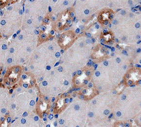 Immunohistochemical analysis of formalin fixed paraffin embedded mouse kidney tissue with F8739 at 1:500 dilution.
Usage Information
| Dilution |
|---|
|
| Application |
|---|
| IHC, IF |
| Reactivity |
|---|
| Mouse |
| Source |
|---|
| Rabbit Monoclonal Antibody |
| Storage Buffer |
|---|
| PBS, pH 7.2+50% Glycerol+0.05% BSA+0.01% NaN3 |
| Storage (from the date of receipt) |
|---|
| -20°C (avoid freeze-thaw cycles), 2 years |
| Predicted MW |
|---|
| 35 kDa |
| Positive Control | Mouse kidney tissue; Mouse liver tissue; RAW 264.7 (LPS, 100 ng/ml, 6 h; BFA, 300 ng/ml, 3 h) |
|---|---|
| Negative Control | RAW 264.7 cells |
Experimental Methods
| IF |
|---|
Experimental Protocol:
Sample Preparation
1. Adherent Cells: Place a clean, sterile coverslip in a culture dish. Once the cells grow to near confluence as a monolayer, remove the coverslip for further use.
2. Suspension Cells: Seed the cells onto a clean, sterile slide coated with poly-L-lysine.
3. Frozen Sections: Allow the slide to thaw at room temperature. Wash it with pure water or PBS for 2 times, 3 minutes each time.
4. Paraffin Sections: Deparaffinization and rehydration. Wash the slide with pure water or PBS for 3 times, 3 minutes each time. Then perform antigen retrieval.
Fixation
1. Fix the cell coverslips/spots or tissue sections at room temperature using a fixative such as 4% paraformaldehyde (4% PFA) for 10-15 minutes.
2. Wash the sample with PBS for 3 times, 3 minutes each time.
Permeabilization
1.Add a detergent such as 0.1–0.3% Triton X-100 to the sample and incubate at room temperature for 10–20 minutes.
(Note: This step is only required for intracellular antigens. For antigens expressed on the cell membrane, this step is unnecessary.)
Wash the sample with PBS for 3 times, 3 minutes each time.
Blocking
Add blocking solution and incubate at room temperature for at least 1 hour. (Common blocking solutions include: serum from the same source as the secondary antibody, BSA, or goat serum.)
Note: Ensure the sample remains moist during and after the blocking step to prevent drying, which can lead to high background.
Immunofluorescence Staining (Day 1)
1. Remove the blocking solution and add the diluted primary antibody.
2. Incubate the sample in a humidified chamber at 4°C overnight.
Immunofluorescence Staining (Day 2)
1. Remove the primary antibody and wash with PBST for 3 times, 5 minutes each time.
2. Add the diluted fluorescent secondary antibody and incubate in the dark at 4°C for 1–2 hours.
3. Remove the secondary antibody and wash with PBST for 3 times, 5 minutes each time.
4. Add diluted DAPI and incubate at room temperature in the dark for 5–10 minutes.
5. Wash with PBST for 3 times, 5 minutes each time.
Mounting
1. Mount the sample with an anti-fade mounting medium.
2. Allow the slide to dry at room temperature overnight in the dark.
3. Store the slide in a slide storage box at 4°C, protected from light.
|
| IHC |
|---|
Experimental Protocol:
Deparaffinization/Rehydration
1. Deparaffinize/hydrate sections:
2. Incubate sections in three washes of xylene for 5 min each.
3. Incubate sections in two washes of 100% ethanol for 10 min each.
4. Incubate sections in two washes of 95% ethanol for 10 min each.
5. Wash sections two times in dH2O for 5 min each.
6.Antigen retrieval: For Citrate: Heat slides in a microwave submersed in 1X citrate unmasking solution until boiling is initiated; continue with 10 min at a sub-boiling temperature (95°-98°C). Cool slides on bench top for 30 min.
Staining
1. Wash sections in dH2O three times for 5 min each.
2. Incubate sections in 3% hydrogen peroxide for 10 min.
3. Wash sections in dH2O two times for 5 min each.
4. Wash sections in wash buffer for 5 min.
5. Block each section with 100–400 µl of blocking solution for 1 hr at room temperature.
6. Remove blocking solution and add 100–400 µl primary antibody diluent in to each section. Incubate overnight at 4°C.
7. Remove antibody solution and wash sections with wash buffer three times for 5 min each.
8. Cover section with 1–3 drops HRPas needed. Incubate in a humidified chamber for 30 min at room temperature.
9. Wash sections three times with wash buffer for 5 min each.
10. Add DAB Chromogen Concentrate to DAB Diluent and mix well before use.
11. Apply 100–400 µl DAB to each section and monitor closely. 1–10 min generally provides an acceptable staining intensity.
12. Immerse slides in dH2O.
13. If desired, counterstain sections with hematoxylin.
14. Wash sections in dH2O two times for 5 min each.
15. Dehydrate sections: Incubate sections in 95% ethanol two times for 10 sec each; Repeat in 100% ethanol, incubating sections two times for 10 sec each; Repeat in xylene, incubating sections two times for 10 sec each.
16. Mount sections with coverslips and mounting medium.
|
Biological Description
| Specificity |
|---|
| Osteopontin Antibody (Rabbit mAb) [G3A21] detects endogenous levels of total Osteopontin protein. |
| Subcellular Location |
|---|
| Secreted |
| Uniprot ID |
|---|
| P10451 |
| Clone |
|---|
| G3A21 |
| Synonym(s) |
|---|
| Eta-1, Op, Spp-1, Spp1, Osteopontin, 2AR, Bone sialoprotein 1, Calcium oxalate crystal growth inhibitor protein, Early T-lymphocyte activation 1 protein, Minopontin, Secreted phosphoprotein 1, SPP-1 |
| Background |
|---|
| Osteopontin, encoded by SPP1, is a highly phosphorylated and glycosylated acidic glycoprotein that functions as both a matrix-associated protein and a secreted cytokine, engaging cell-surface integrins and CD44 through distinct structural motifs. Post-translational modification governs receptor selectivity: phosphorylated osteopontin preferentially binds integrin receptors, while dephosphorylated osteopontin favors CD44 engagement, and O-glycosylation at the C-terminal region further shapes CD44 interaction while also influencing susceptibility to proteolytic cleavage by matrix metalloproteinases. Osteopontin mediates osteoclast attachment to mineralized bone matrix through integrin binding, directly supporting the resorptive activity that governs bone remodeling, and this attachment mechanism positions osteopontin as a physical bridge between the osteoclast cytoskeleton and the bone surface. In prostate cancer cells, osteopontin engagement of integrin αvβ3 activates downstream signaling that promotes CD44 and matrix metalloproteinase-9 complex formation on the cell surface, driving MMP-9 secretion and enzymatic activity that facilitates matrix degradation and cell migration; disrupting the integrin-binding RGD motif of osteopontin or silencing its expression suppresses this CD44-MMP-9 complex formation and reduces migratory capacity. Osteopontin phosphorylation additionally allows the protein to chelate calcium ions through serine and threonine residues, linking its post-translational state to a direct role in regulating hydroxyapatite formation and mineralization dynamics within bone tissue. Serum osteopontin concentrations rise in postmenopausal osteoporosis and correlate inversely with bone mineral density, particularly at the lumbar spine, reflecting the protein's involvement in the resorption-formation imbalance that characterizes the disease. Osteopontin additionally contributes to inflammatory reaction, cell recruitment, and tissue repair processes, and elevated expression across fibrotic, immune-mediated, and malignant tissue contexts links its integrin- and CD44-dependent signaling to disease progression, positioning osteopontin as both a mechanistic driver and a candidate biomarker across bone, inflammatory, and oncologic conditions. |
| References |
|---|
|
Tech Support
Tel: +1-832-582-8158 Ext:3
If you have any other enquiries, please leave a message.

Products are for research use only. Not for human use. We do not sell to patients.
©Copyright 2013 Selleck Chemicals. All Rights Reserved.