-
 Australia
-
 Austria
-
 Belgium
-
 Brazil
-
 Canada
-
 China
China
-
 Czech Republic
-
 Denmark
-
 Finland
-
 France
-
 Germany
-
 Greece
-
 Hong Kong
-
 Hungary
-
 Iceland
-
 India
India
-
 Ireland
-
 Israel
-
 Italy
Italy
-
 Japan
Japan
-
 Korea
Korea
-
 Luxembourg
-
 Malaysia
-
 Netherlands
-
 New Zealand
-
 Norway
-
 Poland
-
 Qatar
Qatar
-
 Romania
-
 Saudi Arabia
-
 Singapore
-
 Spain
Spain
-
 Sweden
-
 Switzerland
-
 Taiwan
-
 Turkey
-
 United Kingdom
-
 United States
research use only
Nurr1 Antibody [L9M11]
Cat.No.: F2094
Application:
Reactivity:
-
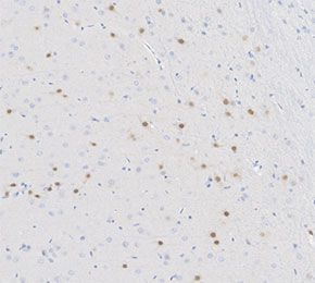 Immunohistochemical analysis of formalin fixed paraffin embedded rat brain tissue with F2094 at 1:100 dilution.
Usage Information
| Dilution |
|---|
|
| Application |
|---|
| IHC |
| Reactivity |
|---|
| Rat |
| Source |
|---|
| Mouse Monoclonal Antibody |
| Storage Buffer |
|---|
| PBS, pH 7.2+50% Glycerol+0.05% BSA+0.01% NaN3 |
| Storage (from the date of receipt) |
|---|
| -20°C (avoid freeze-thaw cycles), 2 years |
| Positive Control | Rat cerebral nerve cell |
|---|---|
| Negative Control |
Experimental Methods
| IHC |
|---|
Experimental Protocol:
Deparaffinization/Rehydration
1. Deparaffinize/hydrate sections:
2. Incubate sections in three washes of xylene for 5 min each.
3. Incubate sections in two washes of 100% ethanol for 10 min each.
4. Incubate sections in two washes of 95% ethanol for 10 min each.
5. Wash sections two times in dH2O for 5 min each.
6.Antigen retrieval: For Citrate: Heat slides in a microwave submersed in 1X citrate unmasking solution until boiling is initiated; continue with 10 min at a sub-boiling temperature (95°-98°C). Cool slides on bench top for 30 min.
Staining
1. Wash sections in dH2O three times for 5 min each.
2. Incubate sections in 3% hydrogen peroxide for 10 min.
3. Wash sections in dH2O two times for 5 min each.
4. Wash sections in wash buffer for 5 min.
5. Block each section with 100–400 µl of blocking solution for 1 hr at room temperature.
6. Remove blocking solution and add 100–400 µl primary antibody diluent in to each section. Incubate overnight at 4°C.
7. Remove antibody solution and wash sections with wash buffer three times for 5 min each.
8. Cover section with 1–3 drops HRPas needed. Incubate in a humidified chamber for 30 min at room temperature.
9. Wash sections three times with wash buffer for 5 min each.
10. Add DAB Chromogen Concentrate to DAB Diluent and mix well before use.
11. Apply 100–400 µl DAB to each section and monitor closely. 1–10 min generally provides an acceptable staining intensity.
12. Immerse slides in dH2O.
13. If desired, counterstain sections with hematoxylin.
14. Wash sections in dH2O two times for 5 min each.
15. Dehydrate sections: Incubate sections in 95% ethanol two times for 10 sec each; Repeat in 100% ethanol, incubating sections two times for 10 sec each; Repeat in xylene, incubating sections two times for 10 sec each.
16. Mount sections with coverslips and mounting medium.
|
Biological Description
| Specificity |
|---|
| Nurr1 Antibody [L9M11] detects endogenous levels of total Nurr1 protein. |
| Subcellular Location |
|---|
| Cytoplasm, Nucleus |
| Uniprot ID |
|---|
| P43354 |
| Clone |
|---|
| L9M11 |
| Synonym(s) |
|---|
| NOT; NURR1; TINUR; NR4A2; Nuclear receptor subfamily 4 group A member 2; Immediate-early response protein NOT; Orphan nuclear receptor NURR1; Transcriptionally-inducible nuclear receptor |
| Background |
|---|
| Nurr1 (NR4A2), an orphan nuclear receptor transcription factor indispensable for midbrain dopaminergic neuron specification, survival, and maintenance, exhibits a modular architecture comprising an N-terminal A/B transactivation domain (AF-1) with phosphorylation sites (Ser336 by ERK), a central DNA-binding domain (DBD) featuring two zinc-finger motifs that recognize NGFI-B response elements (NBRE: AAAGGTCA) as monomers or IR0 half-sites in RXR heterodimers, a flexible hinge region facilitating cofactor recruitment, and a C-terminal ligand-binding domain (LBD) lacking a conventional hydrophobic pocket but exposing a charged helical groove on H11/H12 for coregulator docking via hydrophobic patches. Constitutively active through AF-1/2 synergy, Nurr1 drives dopaminergic gene programs (tyrosine hydroxylase, VMAT2, DAT, Pitx3) by chromatin looping and CBP/p300 coactivation plus RXRα heterodimerization amplifying transcription at composite NBRE-DR5 elements, while post-translational modifications, ERK phosphorylation enhancing AF-1, SUMOylation at K185 repressing via CtBP, GSK3β phosphorylation disrupting RXR interaction, fine-tune activity during development and stress responses, coupling to Wnt/β-catenin for progenitor proliferation and BDNF/TrkB for neuroprotection. Heterozygous mutations (R502X) cause hereditary Parkinsonism with reduced TH+ neurons, while age-related decline exacerbates DA loss in sporadic PD, positioning |
| References |
|---|
|
Tech Support
Tel: +1-832-582-8158 Ext:3
If you have any other enquiries, please leave a message.
Products are for research use only. Not for human use. We do not sell to patients.
©Copyright 2013 Selleck Chemicals. All Rights Reserved.