
-
 Australia
-
 Austria
-
 Belgium
-
 Brazil
-
 Canada
-
 China
China
-
 Czech Republic
-
 Denmark
-
 Finland
-
 France
-
 Germany
-
 Greece
-
 Hong Kong
-
 Hungary
-
 Iceland
-
 India
India
-
 Ireland
-
 Israel
-
 Italy
Italy
-
 Japan
Japan
-
 Korea
Korea
-
 Luxembourg
-
 Malaysia
-
 Netherlands
-
 New Zealand
-
 Norway
-
 Poland
-
 Qatar
Qatar
-
 Romania
-
 Saudi Arabia
-
 Singapore
-
 Spain
Spain
-
 Sweden
-
 Switzerland
-
 Taiwan
-
 Turkey
-
 United Kingdom
-
 United States
research use only
Na Channel β1 Subunit Antibody (Rabbit mAb) [F4D21]
CatNo: F9496
Application:
Reactivity:
-
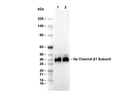 Lane 1: Mouse brain, Lane 2: Rat brain
Experiment Essentials
WB
Recommended wet transfer conditions: 200 mA, 60 min.
Recommended wet transfer conditions: 200 mA, 60 min.
Usage Information
| Dilution |
|---|
|
| Application |
|---|
| WB, IP |
| Reactivity |
|---|
| Human, Mouse, Rat |
| Source |
|---|
| Rabbit Monoclonal Antibody |
| Storage Buffer |
|---|
| PBS, pH 7.2+50% Glycerol+0.05% BSA+0.01% NaN3 |
| Storage (from the date of receipt) |
|---|
| -20°C (avoid freeze-thaw cycles), 2 years |
| Predicted MW |
|---|
| 38 kDa |
| Positive Control | Mouse brain membrane; Rat brain membrane |
|---|---|
| Negative Control |
Experimental Methods
| WB |
|---|
Experimental Protocol:
Sample preparation
1. Tissue: Lyse the tissue sample by adding an appropriate volume of ice-cold RIPA/NP-40 Lysis Buffer (containing Protease Inhibitor Cocktail),and homogenize the tissue at a low temperature or lyse it by sonication on ice, then incubate on ice for 30 minutes. 2. Adherent cell: Aspirate the culture medium and wash the cells with ice-cold PBS twice. Lyse the cells by adding an appropriate volume of RIPA/NP-40 Lysis Buffer (containing Protease Inhibitor Cocktail) , sonicate to lyse the cells, and incubate on ice for 30 minutes. 3. Suspension cell: Transfer the culture medium to a pre-cooled centrifuge tube. Centrifuge and aspirate the supernatant. Wash the cells with ice-cold PBS twice. Lyse the cells by adding an appropriate volume of RIPA/NP-40 Lysis Buffer (containing Protease Inhibitor Cocktail) , sonicate to lyse the cells, and incubate on ice for 30 minutes. 4. Place the lysate into a pre-cooled microcentrifuge tube. Centrifuge at 4°C for 15 min. Collect the supernatant;
5. Remove a small volume of lysate to determine the protein concentration;
6. Combine the lysate with protein loading buffer. Boil 20 µL sample under 95-100°C for 5 min. Centrifuge for 5 min after cool down on ice.
Electrophoretic separation
1. According to the concentration of extracted protein, load appropriate amount of protein sample and marker onto SDS-PAGE gels for electrophoresis. Recommended separating gel (lower gel) concentration: 10%. Reference Table for Selecting SDS-PAGE Separation Gel Concentrations 2. Power up 80V for 30 minutes. Then the power supply is adjusted (110 V~150 V), the Marker is observed, and the electrophoresis can be stopped when the indicator band of the predyed protein Marker where the protein is located is properly separated. (Note that the current should not be too large when electrophoresis, too large current (more than 150 mA) will cause the temperature to rise, affecting the result of running glue. If high currents cannot be avoided, an ice bath can be used to cool the bath.)
Transfer membrane
1. Take out the converter, soak the clip and consumables in the pre-cooled converter;
2. Activate PVDF membrane with methanol for 1 min and rinse with transfer buffer;
3. Install it in the order of "black edge of clip - sponge - filter paper - filter paper - glue -PVDF membrane - filter paper - filter paper - sponge - white edge of clip"; 4. The protein was electrotransferred to PVDF membrane. ( 0.45 µm PVDF membrane is recommended ) Reference Table for Selecting PVDF Membrane Pore Size Specifications Recommended conditions for wet transfer: 200 mA, 60 min. ( Note that the transfer conditions can be adjusted according to the protein size. For high-molecular-weight proteins, a higher current and longer transfer time are recommended. However, ensure that the transfer tank remains at a low temperature to prevent gel melting.)
Block
1. After electrotransfer, wash the film with TBST at room temperature for 5 minutes;
2. Incubate the film in the blocking solution for 1 hour at room temperature;
3. Wash the film with TBST for 3 times, 5 minutes each time.
Antibody incubation
1. Use 5% skim milk powder to prepare the primary antibody working liquid (recommended dilution ratio for primary antibody 1:1000), gently shake and incubate with the film at 4°C overnight; 2. Wash the film with TBST 3 times, 5 minutes each time;
3. Add the secondary antibody to the blocking solution and incubate with the film gently at room temperature for 1 hour;
4. After incubation, wash the film with TBST 3 times for 5 minutes each time.
Antibody staining
1. Add the prepared ECL luminescent substrate (or select other color developing substrate according to the second antibody) and mix evenly;
2. Incubate with the film for 1 minute, remove excess substrate (keep the film moist), wrap with plastic film, and expose in the imaging system. |
Biological Description
| Specificity |
|---|
| Na Channel β1 Subunit Antibody (Rabbit mAb) [F4D21] detects endogenous levels of total Na Channel β1 Subunit protein. |
| Subcellular Location |
|---|
| Cell membrane, Cell projection, Membrane, Secreted |
| Uniprot ID |
|---|
| Q07699 |
| Clone |
|---|
| F4D21 |
| Synonym(s) |
|---|
| Sodium channel regulatory subunit beta-1, SCN1B |
| Background |
|---|
| Na⁺ channel β1 (SCN1B) is a non‑pore‑forming subunit of voltage‑gated sodium channels that combines an extracellular immunoglobulin‑like domain, a single transmembrane segment, and a short intracellular tail, and acts both as a modulator of Nav α‑subunit function and as a cell‑adhesion molecule in excitable tissues. Association of β1 with multiple Nav α‑subunits, including Nav1.1, Nav1.2, Nav1.5, and others, alters channel gating and surface density by shifting activation and inactivation properties, regulating recovery from inactivation, and promoting trafficking of α‑subunits to the plasma membrane, which changes action potential threshold, firing frequency, and conduction in neurons and cardiomyocytes. The extracellular Ig‑like domain supports homophilic and heterophilic cell–cell and cell–matrix adhesion, participates in neurite outgrowth and pathfinding, and localizes Nav complexes to specialized membrane microdomains such as axon initial segments, nodes of Ranvier, and intercalated discs, where coordinated electrical signaling is required. β1 is a substrate for regulated intramembrane proteolysis: sequential cleavage by BACE1 and γ‑secretase generates an intracellular domain that translocates to the nucleus, and transcriptomic analyses identify sets of genes whose expression depends on β1‑ICD levels, linking this fragment to transcriptional control of excitability‑related genes including those encoding Nav1.5. Loss‑of‑function variants in SCN1B reduce β1‑mediated modulation of Nav channels and remove β1‑ICD‑dependent transcriptional restraint, and are associated with developmental and epileptic encephalopathy, generalized epilepsy with febrile seizures plus, and a spectrum of focal and generalized epilepsies that carry high risk of sudden death. SCN1B pathogenic variants are also linked to Brugada syndrome, familial atrial fibrillation, and broader cardiac conduction defects, and mouse models with Scn1b deletion show severe seizures, ventricular arrhythmias, sinoatrial node dysfunction, and atrial remodeling, indicating that β1 contributes to coordinated regulation of neuronal and cardiac excitability. |
| References |
|---|
|
Tech Support
Tel: +1-832-582-8158 Ext:3
If you have any other enquiries, please leave a message.

Products are for research use only. Not for human use. We do not sell to patients.
©Copyright 2013 Selleck Chemicals. All Rights Reserved.