-
 Australia
-
 Austria
-
 Belgium
-
 Brazil
-
 Canada
-
 China
China
-
 Czech Republic
-
 Denmark
-
 Finland
-
 France
-
 Germany
-
 Greece
-
 Hong Kong
-
 Hungary
-
 Iceland
-
 India
India
-
 Ireland
-
 Israel
-
 Italy
Italy
-
 Japan
Japan
-
 Korea
Korea
-
 Luxembourg
-
 Malaysia
-
 Netherlands
-
 New Zealand
-
 Norway
-
 Poland
-
 Qatar
Qatar
-
 Romania
-
 Saudi Arabia
-
 Singapore
-
 Spain
Spain
-
 Sweden
-
 Switzerland
-
 Taiwan
-
 Turkey
-
 United Kingdom
-
 United States
research use only
Myosin light chain kinase/MLCK Antibody (Rabbit mAb) [F16B16]
CatNo: F2230
Application:
Reactivity:
-
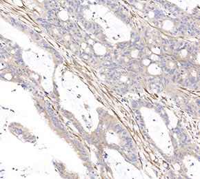 Immunohistochemical analysis of formalin fixed paraffin embedded human colon tissue with F2230 at 1:25000 dilution.
Usage Information
| Dilution |
|---|
|
| Application |
|---|
| IHC |
| Reactivity |
|---|
| Human, Mouse, Rat |
| Source |
|---|
| Rabbit Monoclonal Antibody |
| Storage Buffer |
|---|
| PBS, pH 7.2+50% Glycerol+0.05% BSA+0.01% NaN3 |
| Storage (from the date of receipt) |
|---|
| -20°C (avoid freeze-thaw cycles), 2 years |
| Predicted MW |
|---|
| 211 kDa |
| Positive Control | Human colon tissue; Rat colon tissue; Mouse colon tissue |
|---|---|
| Negative Control |
Experimental Methods
| IHC |
|---|
Experimental Protocol:
Deparaffinization/Rehydration
1. Deparaffinize/hydrate sections:
2. Incubate sections in three washes of xylene for 5 min each.
3. Incubate sections in two washes of 100% ethanol for 10 min each.
4. Incubate sections in two washes of 95% ethanol for 10 min each.
5. Wash sections two times in dH2O for 5 min each.
6.Antigen retrieval: For Citrate: Heat slides in a microwave submersed in 1X citrate unmasking solution until boiling is initiated; continue with 10 min at a sub-boiling temperature (95°-98°C). Cool slides on bench top for 30 min.
Staining
1. Wash sections in dH2O three times for 5 min each.
2. Incubate sections in 3% hydrogen peroxide for 10 min.
3. Wash sections in dH2O two times for 5 min each.
4. Wash sections in wash buffer for 5 min.
5. Block each section with 100–400 µl of blocking solution for 1 hr at room temperature.
6. Remove blocking solution and add 100–400 µl primary antibody diluent in to each section. Incubate overnight at 4°C.
7. Remove antibody solution and wash sections with wash buffer three times for 5 min each.
8. Cover section with 1–3 drops HRPas needed. Incubate in a humidified chamber for 30 min at room temperature.
9. Wash sections three times with wash buffer for 5 min each.
10. Add DAB Chromogen Concentrate to DAB Diluent and mix well before use.
11. Apply 100–400 µl DAB to each section and monitor closely. 1–10 min generally provides an acceptable staining intensity.
12. Immerse slides in dH2O.
13. If desired, counterstain sections with hematoxylin.
14. Wash sections in dH2O two times for 5 min each.
15. Dehydrate sections: Incubate sections in 95% ethanol two times for 10 sec each; Repeat in 100% ethanol, incubating sections two times for 10 sec each; Repeat in xylene, incubating sections two times for 10 sec each.
16. Mount sections with coverslips and mounting medium.
|
Biological Description
| Specificity |
|---|
| Myosin light chain kinase/MLCK Antibody (Rabbit mAb) [F16B16] detects endogenous levels of total Myosin light chain kinase/MLCK protein. |
| Uniprot ID |
|---|
| Q15746 |
| Clone |
|---|
| F16B16 |
| Synonym(s) |
|---|
| MLCK, MLCK1, MYLK1, MYLK, smMLCK, Kinase-related protein, Telokin, KRP |
| Background |
|---|
| Myosin light chain kinase (MLCK, MYLK) is a Ca²⁺/calmodulin-dependent serine/threonine kinase of the myosin kinase family that phosphorylates the regulatory light chain of myosin II and couples Ca²⁺ signals to actomyosin contractility in smooth muscle, skeletal muscle, and nonmuscle cells. The enzyme contains an N‑terminal targeting region that associates with actin–myosin filaments, a central catalytic kinase domain with the consensus HRD motif, and a C‑terminal regulatory segment harboring the calmodulin-binding site and multiple regulatory phosphorylation sites, and this arrangement allows Ca²⁺/calmodulin binding and upstream kinase inputs to control catalytic activity and subcellular positioning. Activation begins when Ca²⁺ binds calmodulin, and the Ca²⁺/calmodulin complex engages the MLCK regulatory domain, relieving autoinhibition and enabling MLCK to phosphorylate the regulatory myosin light chain at a conserved serine, which increases myosin ATPase activity, promotes actin binding, and initiates cross-bridge cycling and force generation in smooth muscle or potentiates force and shortening in skeletal muscle. Smooth muscle–specific MLCK isoforms are further tuned by phosphorylation of residues within the calmodulin-binding region by kinases such as CaMKII, PKA, PKG, PAK, and PKC, which reduce calmodulin affinity and thereby decrease sensitivity to Ca²⁺, while mitogen-activated protein kinases phosphorylate other sites to increase maximal catalytic velocity without changing Ca²⁺ dependence, integrating diverse signaling pathways onto the same contractile effector. MLCK operates in dynamic balance with myosin light chain phosphatase, and the relative activities of these enzymes define the phosphorylation state of myosin light chain, the level of actomyosin interaction, and the presence of latch-like contractile states in smooth muscle. Nonmuscle MLCK variants localize to cortical and junctional actomyosin networks, where they regulate stress fiber organization, focal adhesion maturation, and perijunctional actomyosin ring contraction, linking integrin and Rho family GTPase signaling to cell shape, adhesion, and migration. In endothelial cells, MLCK-dependent phosphorylation of myosin light chain at the cell borders drives contraction of the apical actomyosin ring, increases paracellular gap formation, and modulates microvascular barrier permeability during inflammatory responses, ischemia–reperfusion, and cardiovascular stress. Abnormal MLCK expression or activity is associated with inflammatory diseases, acute lung injury, asthma, and other conditions where exaggerated actomyosin contraction and barrier dysfunction promote edema and leukocyte recruitment, and pharmacologic MLCK inhibition reduces myosin light chain phosphorylation, stabilizes junctions, and attenuates inflammatory leakage in experimental models. |
| References |
|---|
|
Tech Support
Tel: +1-832-582-8158 Ext:3
If you have any other enquiries, please leave a message.
Products are for research use only. Not for human use. We do not sell to patients.
©Copyright 2013 Selleck Chemicals. All Rights Reserved.