-
 Australia
-
 Austria
-
 Belgium
-
 Brazil
-
 Canada
-
 China
China
-
 Czech Republic
-
 Denmark
-
 Finland
-
 France
-
 Germany
-
 Greece
-
 Hong Kong
-
 Hungary
-
 Iceland
-
 India
India
-
 Ireland
-
 Israel
-
 Italy
Italy
-
 Japan
Japan
-
 Korea
Korea
-
 Luxembourg
-
 Malaysia
-
 Netherlands
-
 New Zealand
-
 Norway
-
 Poland
-
 Qatar
Qatar
-
 Romania
-
 Saudi Arabia
-
 Singapore
-
 Spain
Spain
-
 Sweden
-
 Switzerland
-
 Taiwan
-
 Turkey
-
 United Kingdom
-
 United States
research use only
MAD2L2 Antibody [P22A23]
Cat.No.: F5275
Application:
Reactivity:
-
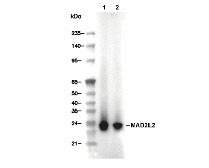 Lane 1: NCI-H1963, Lane 2: NCCIT
Experiment Essentials
WB
Recommended wet transfer conditions: 200 mA, 60 min.
Recommended wet transfer conditions: 200 mA, 60 min.
Usage Information
| Dilution |
|---|
|
| Application |
|---|
| WB, IP |
| Reactivity |
|---|
| Human, Mouse, Rat |
| Source |
|---|
| Rabbit Monoclonal Antibody |
| Storage Buffer |
|---|
| PBS, pH 7.2+50% Glycerol+0.05% BSA+0.01% NaN3 |
| Storage (from the date of receipt) |
|---|
| -20°C (avoid freeze-thaw cycles), 2 years |
| Predicted MW |
|---|
| 24 kDa |
| Positive Control | NCI-H1963 cells; NCCIT cells; Calu cells; NMuMG cells; MBT2 cells; MTLn3 cells |
|---|---|
| Negative Control |
Experimental Methods
| WB |
|---|
Experimental Protocol:
Sample preparation
1. Tissue: Lyse the tissue sample by adding an appropriate volume of ice-cold RIPA/NP-40 Lysis Buffer (containing Protease Inhibitor Cocktail),and homogenize the tissue at a low temperature. 2. Adherent cell: Aspirate the culture medium and wash the cells with ice-cold PBS twice. Lyse the cells by adding an appropriate volume of RIPA/NP-40 Lysis Buffer (containing Protease Inhibitor Cocktail) and put the sample on ice for 5 min. 3. Suspension cell: Transfer the culture medium to a pre-cooled centrifuge tube. Centrifuge and aspirate the supernatant. Wash the cells with ice-cold PBS twice. Lyse the cells by adding an appropriate volume of RIPA/NP-40 Lysis Buffer (containing Protease Inhibitor Cocktail) and put the sample on ice for 5 min. 4. Place the lysate into a pre-cooled microcentrifuge tube. Centrifuge at 4°C for 15 min. Collect the supernatant;
5. Remove a small volume of lysate to determine the protein concentration;
6. Combine the lysate with protein loading buffer. Boil 20 µL sample under 95-100°C for 5 min. Centrifuge for 5 min after cool down on ice.
Electrophoretic separation
1. According to the concentration of extracted protein, load appropriate amount of protein sample and marker onto SDS-PAGE gels for electrophoresis. Recommended separating gel (lower gel) concentration: 10%. Reference Table for Selecting SDS-PAGE Separation Gel Concentrations 2. Power up 80V for 30 minutes. Then the power supply is adjusted (110 V~150 V), the Marker is observed, and the electrophoresis can be stopped when the indicator band of the predyed protein Marker where the protein is located is properly separated. (Note that the current should not be too large when electrophoresis, too large current (more than 150 mA) will cause the temperature to rise, affecting the result of running glue. If high currents cannot be avoided, an ice bath can be used to cool the bath.)
Transfer membrane
1. Take out the converter, soak the clip and consumables in the pre-cooled converter;
2. Activate PVDF membrane with methanol for 1 min and rinse with transfer buffer;
3. Install it in the order of "black edge of clip - sponge - filter paper - filter paper - glue -PVDF membrane - filter paper - filter paper - sponge - white edge of clip"; 4. The protein was electrotransferred to PVDF membrane. ( 0.45 µm PVDF membrane is recommended ) Reference Table for Selecting PVDF Membrane Pore Size Specifications Recommended conditions for wet transfer: 200 mA, 60 min. ( Note that the transfer conditions can be adjusted according to the protein size. For high-molecular-weight proteins, a higher current and longer transfer time are recommended. However, ensure that the transfer tank remains at a low temperature to prevent gel melting.)
Block
1. After electrotransfer, wash the film with TBST at room temperature for 5 minutes;
2. Incubate the film in the blocking solution for 1 hour at room temperature;
3. Wash the film with TBST for 3 times, 5 minutes each time.
Antibody incubation
1. Use 5% skim milk powder to prepare the primary antibody working liquid (recommended dilution ratio for primary antibody 1:1000), gently shake and incubate with the film at 4°C overnight; 2. Wash the film with TBST 3 times, 5 minutes each time;
3. Add the secondary antibody to the blocking solution and incubate with the film gently at room temperature for 1 hour;
4. After incubation, wash the film with TBST 3 times for 5 minutes each time.
Antibody staining
1. Add the prepared ECL luminescent substrate (or select other color developing substrate according to the second antibody) and mix evenly;
2. Incubate with the film for 1 minute, remove excess substrate (keep the film moist), wrap with plastic film, and expose in the imaging system. |
Biological Description
| Specificity |
|---|
| MAD2L2 Antibody [P22A23] detects endogenous levels of total MAD2L2 protein. |
| Subcellular Location |
|---|
| Chromosome, Cytoplasm, Cytoskeleton, Nucleus |
| Uniprot ID |
|---|
| Q9UI95 |
| Clone |
|---|
| P22A23 |
| Synonym(s) |
|---|
| Mitotic spindle assembly checkpoint protein MAD2B; MAD2-like protein 2; REV7; MAD2L2 |
| Background |
|---|
| Mitotic arrest deficient 2 like 2 (MAD2L2, also known as REV7) is a multifunctional HORMA‑domain protein that integrates cell‑cycle control with DNA damage response by participating in the spindle checkpoint machinery and several DNA repair pathways. It belongs to the MAD2‑like or HORMA family and adopts a closing‑clamshell architecture that enables it to act as an adaptor or scaffold, engaging partner proteins through interface‑switching modes rather than forming the core of the spindle assembly checkpoint itself. MAD2L2 functions as a core subunit of DNA polymerase ζ, where it bridges the catalytic subunit REV3 and the TLS scaffold REV1, coordinating lesion bypass during translesion synthesis and enabling replication through blocking DNA adducts. In double‑strand break repair, MAD2L2 joins REV1 and polymerase ζ at stalled forks and contributes to repair‑synthesis steps, while also participating in the shieldin complex, where it helps regulate pathway choice by limiting 5ʹ end resection and thereby favoring non‑homologous end joining over homologous recombination. MAD2L2 also contributes to mitotic regulation by restraining premature activation of the anaphase‑promoting complex/cyclosome through interactions with CDH1 and related checkpoint proteins, thereby helping to delay mitotic progression and maintain genome integrity under replication stress. MAD2L2 is frequently overexpressed compared with adjacent normal tissue, and high message or protein levels correlate with increased numbers of aberrant mitotic figures, chromosomal instability, and reduced overall survival in colorectal cancer and other solid malignancies, suggesting that elevated MAD2L2 expression reinforces error‑prone DNA repair and checkpoint adaptations that support tumor evolution. |
| References |
|---|
|
Tech Support
Tel: +1-832-582-8158 Ext:3
If you have any other enquiries, please leave a message.
Products are for research use only. Not for human use. We do not sell to patients.
©Copyright 2013 Selleck Chemicals. All Rights Reserved.