-
 Australia
-
 Austria
-
 Belgium
-
 Brazil
-
 Canada
-
 China
China
-
 Czech Republic
-
 Denmark
-
 Finland
-
 France
-
 Germany
-
 Greece
-
 Hong Kong
-
 Hungary
-
 Iceland
-
 India
India
-
 Ireland
-
 Israel
-
 Italy
Italy
-
 Japan
Japan
-
 Korea
Korea
-
 Luxembourg
-
 Malaysia
-
 Netherlands
-
 New Zealand
-
 Norway
-
 Poland
-
 Qatar
Qatar
-
 Romania
-
 Saudi Arabia
-
 Singapore
-
 Spain
Spain
-
 Sweden
-
 Switzerland
-
 Taiwan
-
 Turkey
-
 United Kingdom
-
 United States
research use only
Ghrelin Antibody [F2E5]
Cat.No.: F3727
Application:
Reactivity:
-
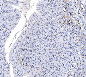 Immunohistochemical analysis of formalin fixed paraffin embedded mouse stomach tissue with F3727 at 1:8000 dilution.
Usage Information
| Dilution |
|---|
|
| Application |
|---|
| IHC |
| Reactivity |
|---|
| Mouse, Rat, Human |
| Source |
|---|
| Rabbit Monoclonal Antibody |
| Storage Buffer |
|---|
| PBS, pH 7.2+50% Glycerol+0.05% BSA+0.01% NaN3 |
| Storage (from the date of receipt) |
|---|
| -20°C (avoid freeze-thaw cycles), 2 years |
| Positive Control | Mouse stomach tissue; Human stomach tissue; Rat stomach tissue |
|---|---|
| Negative Control | Rat liver tissue; Mouse liver tissue; Human liver tissue |
Experimental Methods
| IHC |
|---|
Experimental Protocol:
Deparaffinization/Rehydration
1. Deparaffinize/hydrate sections:
2. Incubate sections in three washes of xylene for 5 min each.
3. Incubate sections in two washes of 100% ethanol for 10 min each.
4. Incubate sections in two washes of 95% ethanol for 10 min each.
5. Wash sections two times in dH2O for 5 min each.
6.Antigen retrieval: For Citrate: Heat slides in a microwave submersed in 1X citrate unmasking solution until boiling is initiated; continue with 10 min at a sub-boiling temperature (95°-98°C). Cool slides on bench top for 30 min.
Staining
1. Wash sections in dH2O three times for 5 min each.
2. Incubate sections in 3% hydrogen peroxide for 10 min.
3. Wash sections in dH2O two times for 5 min each.
4. Wash sections in wash buffer for 5 min.
5. Block each section with 100–400 µl of blocking solution for 1 hr at room temperature.
6. Remove blocking solution and add 100–400 µl primary antibody diluent in to each section. Incubate overnight at 4°C.
7. Remove antibody solution and wash sections with wash buffer three times for 5 min each.
8. Cover section with 1–3 drops HRPas needed. Incubate in a humidified chamber for 30 min at room temperature.
9. Wash sections three times with wash buffer for 5 min each.
10. Add DAB Chromogen Concentrate to DAB Diluent and mix well before use.
11. Apply 100–400 µl DAB to each section and monitor closely. 1–10 min generally provides an acceptable staining intensity.
12. Immerse slides in dH2O.
13. If desired, counterstain sections with hematoxylin.
14. Wash sections in dH2O two times for 5 min each.
15. Dehydrate sections: Incubate sections in 95% ethanol two times for 10 sec each; Repeat in 100% ethanol, incubating sections two times for 10 sec each; Repeat in xylene, incubating sections two times for 10 sec each.
16. Mount sections with coverslips and mounting medium.
|
Biological Description
| Specificity |
|---|
| Ghrelin Antibody [F2E5] detects endogenous levels of total Ghrelin protein. |
| Subcellular Location |
|---|
| Secreted |
| Uniprot ID |
|---|
| Q9UBU3 |
| Clone |
|---|
| F2E5 |
| Synonym(s) |
|---|
| MTLRP; UNQ524/PRO1066; GHRL; Appetite‑regulating hormone; Growth hormone secretagogue; Growth hormone‑releasing peptide; Motilin‑related peptide; Protein M46 |
| Background |
|---|
| Ghrelin is an acylated peptide hormone synthesized predominantly in gastric enteroendocrine cells, with additional expression in the pancreas, lung, and hypothalamic neurons, where it acts as a key peripheral and central regulator of energy homeostasis, growth hormone release, and reward related feeding behavior. The mature ligand binds to the growth hormone secretagogue receptor (GHSR/GHS R1a), a class A G protein–coupled receptor highly expressed in the hypothalamic arcuate nucleus, pituitary, and midbrain dopamine circuits, engaging multiple G protein families including Gαq/11, Gαi/o, and Gα12/13, which activate phospholipase C–inositol trisphosphate–calcium signaling, inhibit adenylate cyclase, and recruit Rho A–dependent pathways, respectively, while β arrestins additionally mediate GHSR internalization and MAPK activation. In the arcuate nucleus, ghrelin GHSR signaling stimulates neuropeptide Y and agouti related peptide–expressing orexigenic neurons, increases AMP activated protein kinase activity, and disinhibits dopaminergic reward pathways, thereby promoting hunger, anticipatory feeding, and preference for calorie dense foods, whereas in the pituitary the same receptor ligand pair drives calcium dependent growth hormone secretion, linking ghrelin to somatotropic axis control and metabolic adaptation. Ghrelin also modulates gastric motility, acid secretion, and pancreatic islet function. Ghrelin-mediated Gαi signalling attenuates glucose-stimulated insulin release, thereby coordinating energy intake, storage, and fuel utilisation under fasted states. In contrast, its circulating levels normally decline after meals to disinhibit satiety pathways governed by leptin and insulin. In obesity, ghrelin suppression can blunt meal-induced inhibition of appetite and impair metabolic flexibility. Conversely, in cachexia and chronic illness, reduced ghrelin tone may exacerbate negative energy balance and muscle wasting, ultimately leading to dysregulated ghrelin-GHSR signalling across both hyperphagic and catabolic metabolic states. |
| References |
|---|
|
Tech Support
Tel: +1-832-582-8158 Ext:3
If you have any other enquiries, please leave a message.
Products are for research use only. Not for human use. We do not sell to patients.
©Copyright 2013 Selleck Chemicals. All Rights Reserved.