
-
 Australia
-
 Austria
-
 Belgium
-
 Brazil
-
 Canada
-
 China
China
-
 Czech Republic
-
 Denmark
-
 Finland
-
 France
-
 Germany
-
 Greece
-
 Hong Kong
-
 Hungary
-
 Iceland
-
 India
India
-
 Ireland
-
 Israel
-
 Italy
Italy
-
 Japan
Japan
-
 Korea
Korea
-
 Luxembourg
-
 Malaysia
-
 Netherlands
-
 New Zealand
-
 Norway
-
 Poland
-
 Qatar
Qatar
-
 Romania
-
 Saudi Arabia
-
 Singapore
-
 Spain
Spain
-
 Sweden
-
 Switzerland
-
 Taiwan
-
 Turkey
-
 United Kingdom
-
 United States
research use only
Galectin 8/Gal-8 Antibody (Rabbit mAb) [J21J20]
CatNo: F7782
Application:
Reactivity:
-
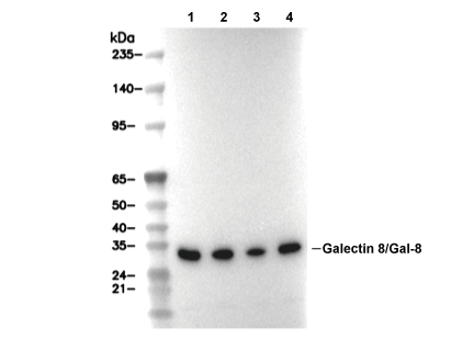 Lane 1: RAW 264.7, Lane 2: C2C12, Lane 3: Mouse brain, Lane 4: Rat brain
Experiment Essentials
WB
Recommended wet transfer conditions: 200 mA, 60 min.
Recommended wet transfer conditions: 200 mA, 60 min.
Usage Information
| Dilution |
|---|
|
| Application |
|---|
| WB |
| Reactivity |
|---|
| Mouse, Rat |
| Source |
|---|
| Rabbit Monoclonal Antibody |
| Storage Buffer |
|---|
| PBS, pH 7.2+50% Glycerol+0.05% BSA+0.01% NaN3 |
| Storage (from the date of receipt) |
|---|
| -20°C (avoid freeze-thaw cycles), 2 years |
| Predicted MW Observed MW |
|---|
| 36 kDa 34 kDa,36 kDa |
| *Why do the predicted and actual molecular weights differ? The following reasons may explain differences between the predicted and actual protein molecular weight. Post-translational modifications(e.g., phosphorylation, glycosylation); Splice variants and isoforms; Relative charge; Multimerization. |
| Positive Control | Rat brain tissue; Rat heart tissue; Rat spleen tissue; Mouse brain tissue; Mouse heart tissue; Mouse spleen tissue; RAW 264.7 cells; C2C12 cells; PC-12 cells; C6 cells |
|---|---|
| Negative Control |
Experimental Methods
| WB |
|---|
Experimental Protocol:
Sample preparation
1. Tissue: Lyse the tissue sample by adding an appropriate volume of ice-cold RIPA/NP-40 Lysis Buffer (containing Protease Inhibitor Cocktail),and homogenize the tissue at a low temperature or lyse it by sonication on ice, then incubate on ice for 30 minutes. 2. Adherent cell: Aspirate the culture medium and wash the cells with ice-cold PBS twice. Lyse the cells by adding an appropriate volume of RIPA/NP-40 Lysis Buffer (containing Protease Inhibitor Cocktail) , sonicate to lyse the cells, and incubate on ice for 30 minutes. 3. Suspension cell: Transfer the culture medium to a pre-cooled centrifuge tube. Centrifuge and aspirate the supernatant. Wash the cells with ice-cold PBS twice. Lyse the cells by adding an appropriate volume of RIPA/NP-40 Lysis Buffer (containing Protease Inhibitor Cocktail) , sonicate to lyse the cells, and incubate on ice for 30 minutes. 4. Place the lysate into a pre-cooled microcentrifuge tube. Centrifuge at 4°C for 15 min. Collect the supernatant;
5. Remove a small volume of lysate to determine the protein concentration;
6. Combine the lysate with protein loading buffer. Boil 20 µL sample under 95-100°C for 5 min. Centrifuge for 5 min after cool down on ice.
Electrophoretic separation
1. According to the concentration of extracted protein, load appropriate amount of protein sample and marker onto SDS-PAGE gels for electrophoresis. Recommended separating gel (lower gel) concentration: 10%. Reference Table for Selecting SDS-PAGE Separation Gel Concentrations 2. Power up 80V for 30 minutes. Then the power supply is adjusted (110 V~150 V), the Marker is observed, and the electrophoresis can be stopped when the indicator band of the predyed protein Marker where the protein is located is properly separated. (Note that the current should not be too large when electrophoresis, too large current (more than 150 mA) will cause the temperature to rise, affecting the result of running glue. If high currents cannot be avoided, an ice bath can be used to cool the bath.)
Transfer membrane
1. Take out the converter, soak the clip and consumables in the pre-cooled converter;
2. Activate PVDF membrane with methanol for 1 min and rinse with transfer buffer;
3. Install it in the order of "black edge of clip - sponge - filter paper - filter paper - glue -PVDF membrane - filter paper - filter paper - sponge - white edge of clip"; 4. The protein was electrotransferred to PVDF membrane. ( 0.45 µm PVDF membrane is recommended ) Reference Table for Selecting PVDF Membrane Pore Size Specifications Recommended conditions for wet transfer: 200 mA, 60 min. ( Note that the transfer conditions can be adjusted according to the protein size. For high-molecular-weight proteins, a higher current and longer transfer time are recommended. However, ensure that the transfer tank remains at a low temperature to prevent gel melting.)
Block
1. After electrotransfer, wash the film with TBST at room temperature for 5 minutes;
2. Incubate the film in the blocking solution for 1 hour at room temperature;
3. Wash the film with TBST for 3 times, 5 minutes each time.
Antibody incubation
1. Use primary antibody dilution buffer to prepare the primary antibody working liquid (recommended dilution ratio for primary antibody 1:1000), gently shake and incubate with the film at 4°C overnight; 2. Wash the film with TBST 3 times, 5 minutes each time;
3. Add the secondary antibody to the blocking solution and incubate with the film gently at room temperature for 1 hour;
4. After incubation, wash the film with TBST 3 times for 5 minutes each time.
Antibody staining
1. Add the prepared ECL luminescent substrate (or select other color developing substrate according to the second antibody) and mix evenly;
2. Incubate with the film for 1 minute, remove excess substrate (keep the film moist), wrap with plastic film, and expose in the imaging system. |
Biological Description
| Specificity |
|---|
| Galectin 8/Gal-8 Antibody (Rabbit mAb) [J21J20] detects endogenous levels of total Galectin 8/Gal-8 protein. |
| Subcellular Location |
|---|
| Cytoplasm, Cytoplasmic vesicle |
| Uniprot ID |
|---|
| O00214 |
| Clone |
|---|
| J21J20 |
| Synonym(s) |
|---|
| Galectin-8, Gal-8, LGALS-8, Lgals8 |
| Background |
|---|
| Galectin-8 belongs to the tandem-repeat subfamily of the galectin family of beta-galactoside-binding lectins, distinguished structurally from prototype galectins by possessing two distinct carbohydrate recognition domains, an N-terminal domain and a C-terminal domain, connected by a linker peptide rather than a single self-dimerizing binding domain. These two domains carry markedly different glycan-binding specificities: the N-terminal domain shows strong preferential affinity for alpha2-3-sialylated and 3'-sulfated beta-galactosides, while the C-terminal domain instead favors non-sialylated oligosaccharide structures, including poly-N-acetyllactosamine and blood group A and B antigen glycans, giving a single Galectin-8 molecule two functionally distinct recognition surfaces capable of engaging different classes of cell-surface and intracellular glycoconjugates simultaneously. Functioning as a cytosolic danger receptor, Galectin-8 monitors the integrity of endosomal and lysosomal membranes by detecting host glycans that become abnormally exposed to the cytosol when these membranes are ruptured, whether by invading bacteria such as Salmonella, Listeria, or Shigella, or by sterile membrane damage; because complex glycans are normally confined to the luminal, extracellular-facing side of vesicular membranes, their sudden cytosolic exposure functions as an unambiguous signal of membrane rupture that Galectin-8 is positioned to recognize directly. Upon binding these exposed glycans on a damaged, bacteria-containing vacuole, Galectin-8 recruits the autophagy receptor NDP52 to the site, and this Galectin-8-dependent NDP52 recruitment is transient and precedes a subsequent, ubiquitin-dependent phase of NDP52 recruitment, indicating that Galectin-8 initiates antibacterial autophagy through a glycan-sensing mechanism that operates upstream of and independently from the classical polyubiquitin-coating route used to flag pathogens for autophagic clearance. Galectin-8 recruitment to damaged vesicles occurs independently of core autophagy machinery, since it proceeds normally even in cells lacking ATG5, confirming that Galectin-8 functions as a genuine pattern-recognition sensor of vesicle damage rather than as a downstream component of the autophagy pathway itself. Beyond this innate immune, danger-sensing function, Galectin-8 engages numerous cell-surface glycoprotein ligands through its two structurally distinct CRDs, including integrins, CD44, CD45, and basigin/CD147, mediating cell adhesion, immune cell regulation, and modulation of T-lymphocyte activity, and elevated LGALS8 expression across a broad range of human tumors, together with its established role in promoting angiogenesis and lymphangiogenesis, has made Galectin-8, and particularly its N-terminal CRD, an actively pursued target for small-molecule inhibitor development in cancer. |
| References |
|---|
|
Tech Support
Tel: +1-832-582-8158 Ext:3
If you have any other enquiries, please leave a message.

Products are for research use only. Not for human use. We do not sell to patients.
©Copyright 2013 Selleck Chemicals. All Rights Reserved.