-
 Australia
-
 Austria
-
 Belgium
-
 Brazil
-
 Canada
-
 China
China
-
 Czech Republic
-
 Denmark
-
 Finland
-
 France
-
 Germany
-
 Greece
-
 Hong Kong
-
 Hungary
-
 Iceland
-
 India
India
-
 Ireland
-
 Israel
-
 Italy
Italy
-
 Japan
Japan
-
 Korea
Korea
-
 Luxembourg
-
 Malaysia
-
 Netherlands
-
 New Zealand
-
 Norway
-
 Poland
-
 Qatar
Qatar
-
 Romania
-
 Saudi Arabia
-
 Singapore
-
 Spain
Spain
-
 Sweden
-
 Switzerland
-
 Taiwan
-
 Turkey
-
 United Kingdom
-
 United States
research use only
FUNDC1 Antibody [A14J2]
Cat.No.: F9508
Application:
Reactivity:
-
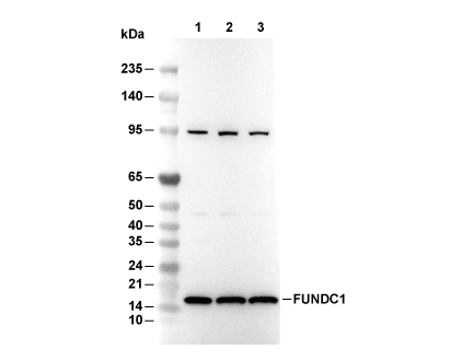 Lane 1: RAW264.7, Lane 2: C6, Lane 3: H-4-II-E
Experiment Essentials
WB
Recommended wet transfer conditions: 200 mA, 60 min,Recommended to use 0.22 μm PVDF membrane.
Recommended wet transfer conditions: 200 mA, 60 min,Recommended to use 0.22 μm PVDF membrane.
Usage Information
| Dilution |
|---|
|
| Application |
|---|
| WB, IP |
| Reactivity |
|---|
| Human, Mouse, Rat |
| Source |
|---|
| Rabbit Monoclonal Antibody |
| Storage Buffer |
|---|
| PBS, pH 7.2+50% Glycerol+0.05% BSA+0.01% NaN3 |
| Storage (from the date of receipt) |
|---|
| -20°C (avoid freeze-thaw cycles), 2 years |
| Predicted MW |
|---|
| 17 kDa |
| Positive Control | A‑673 cells; BT‑483 cells; A2780 cells; HCC1428 cells; ZR‑75‑1 cells; RAW 264.7 cells; JAWSII cells; C6 cells; H‑4‑II‑E cells |
|---|---|
| Negative Control | THP‑1 cells; MIA PaCa‑2 cells |
Experimental Methods
| WB |
|---|
Experimental Protocol:
Sample preparation
1. Tissue: Lyse the tissue sample by adding an appropriate volume of ice-cold RIPA/NP-40 Lysis Buffer (containing Protease Inhibitor Cocktail),and homogenize the tissue at a low temperature. 2. Adherent cell: Aspirate the culture medium and wash the cells with ice-cold PBS twice. Lyse the cells by adding an appropriate volume of RIPA/NP-40 Lysis Buffer (containing Protease Inhibitor Cocktail) and put the sample on ice for 5 min. 3. Suspension cell: Transfer the culture medium to a pre-cooled centrifuge tube. Centrifuge and aspirate the supernatant. Wash the cells with ice-cold PBS twice. Lyse the cells by adding an appropriate volume of RIPA/NP-40 Lysis Buffer (containing Protease Inhibitor Cocktail) and put the sample on ice for 5 min. 4. Place the lysate into a pre-cooled microcentrifuge tube. Centrifuge at 4°C for 15 min. Collect the supernatant;
5. Remove a small volume of lysate to determine the protein concentration;
6. Combine the lysate with protein loading buffer. Boil 20 µL sample under 95-100°C for 5 min. Centrifuge for 5 min after cool down on ice.
Electrophoretic separation
1. According to the concentration of extracted protein, load appropriate amount of protein sample and marker onto SDS-PAGE gels for electrophoresis. Recommended separating gel (lower gel) concentration: 10%. Reference Table for Selecting SDS-PAGE Separation Gel Concentrations 2. Power up 80V for 30 minutes. Then the power supply is adjusted (110 V~150 V), the Marker is observed, and the electrophoresis can be stopped when the indicator band of the predyed protein Marker where the protein is located is properly separated. (Note that the current should not be too large when electrophoresis, too large current (more than 150 mA) will cause the temperature to rise, affecting the result of running glue. If high currents cannot be avoided, an ice bath can be used to cool the bath.)
Transfer membrane
1. Take out the converter, soak the clip and consumables in the pre-cooled converter;
2. Activate PVDF membrane with methanol for 1 min and rinse with transfer buffer;
3. Install it in the order of "black edge of clip - sponge - filter paper - filter paper - glue -PVDF membrane - filter paper - filter paper - sponge - white edge of clip"; 4. The protein was electrotransferred to PVDF membrane. ( 0.22 µm PVDF membrane is recommended )) Reference Table for Selecting PVDF Membrane Pore Size Specifications Recommended conditions for wet transfer: 200 mA, 60 min. ( Note that the transfer conditions can be adjusted according to the protein size. For high-molecular-weight proteins, a higher current and longer transfer time are recommended. However, ensure that the transfer tank remains at a low temperature to prevent gel melting.)
Block
1. After electrotransfer, wash the film with TBST at room temperature for 5 minutes;
2. Incubate the film in the blocking solution for 1 hour at room temperature;
3. Wash the film with TBST for 3 times, 5 minutes each time.
Antibody incubation
1. Use 5% skim milk powder to prepare the primary antibody working liquid (recommended dilution ratio for primary antibody 1:1000), gently shake and incubate with the film at 4°C overnight; 2. Wash the film with TBST 3 times, 5 minutes each time;
3. Add the secondary antibody to the blocking solution and incubate with the film gently at room temperature for 1 hour;
4. After incubation, wash the film with TBST 3 times for 5 minutes each time.
Antibody staining
1. Add the prepared ECL luminescent substrate (or select other color developing substrate according to the second antibody) and mix evenly;
2. Incubate with the film for 1 minute, remove excess substrate (keep the film moist), wrap with plastic film, and expose in the imaging system. |
Biological Description
| Specificity |
|---|
| FUNDC1 Antibody [A14J2] detects endogenous levels of total FUNDC1 protein. |
| Subcellular Location |
|---|
| Membrane, Mitochondrion, Mitochondrion outer membrane |
| Uniprot ID |
|---|
| Q8IVP5 |
| Clone |
|---|
| A14J2 |
| Synonym(s) |
|---|
| FUN14 domain-containing protein 1; FUNDC1 |
| Background |
|---|
| FUNDC1 anchors to the mitochondrial outer membrane as an OMM protein that mediates hypoxia-induced mitophagy through direct interaction with LC3 family proteins on forming autophagosomes. The protein features an N-terminal LIR motif with a core W/F/Y-xx-L/I/V sequence flanked by flanking acidic residues that engage LC3 hydrophobic pockets, alongside conserved phosphorylation sites at tyrosine 18, serine 13, and serine 17 that reversibly modulate binding affinity. Under normoxia, Src kinase phosphorylates Tyr18 to disrupt LIR-LC3 interaction while CK2 targets Ser13 to maintain inhibitory conformation, preventing mitophagosome formation; hypoxia inactivates Src and activates PGAM5 phosphatase to dephosphorylate both sites, exposing the LIR for high-affinity LC3/GABARAP binding and Parkin-independent mitochondrial clearance. ULK1/ATG1 kinase phosphorylates Ser17 during starvation to further enhance LC3 association and autophagosome engulfment, while MARCH5 E3 ligase ubiquitinates lysine residues under steady state to target FUNDC1 for proteasomal degradation via the cytosol-to-mitochondria import pathway. FUNDC1 coordinates with Drp1 to regulate mitochondrial fission at ER-mitochondria contact sites through Tyr18 dephosphorylation, balancing fusion-fission dynamics during metabolic stress. The protein forms a tripartite complex with PGAM5 and CK2α to create a hypoxia-responsive switch that integrates kinase-phosphatase signaling with LC3 lipidation cascades. FUNDC1 deficiency impairs hypoxic adaptation in cardiomyocytes and hepatocytes, leading to mitochondrial accumulation and ROS-mediated damage. Phosphomimetic mutants at Ser13/17 block mitophagy induction, while non-phosphorylatable Tyr18 variants constitutively activate clearance. |
| References |
|---|
|
Tech Support
Tel: +1-832-582-8158 Ext:3
If you have any other enquiries, please leave a message.
Products are for research use only. Not for human use. We do not sell to patients.
©Copyright 2013 Selleck Chemicals. All Rights Reserved.