-
 Australia
-
 Austria
-
 Belgium
-
 Brazil
-
 Canada
-
 China
China
-
 Czech Republic
-
 Denmark
-
 Finland
-
 France
-
 Germany
-
 Greece
-
 Hong Kong
-
 Hungary
-
 Iceland
-
 India
India
-
 Ireland
-
 Israel
-
 Italy
Italy
-
 Japan
Japan
-
 Korea
Korea
-
 Luxembourg
-
 Malaysia
-
 Netherlands
-
 New Zealand
-
 Norway
-
 Poland
-
 Qatar
Qatar
-
 Romania
-
 Saudi Arabia
-
 Singapore
-
 Spain
Spain
-
 Sweden
-
 Switzerland
-
 Taiwan
-
 Turkey
-
 United Kingdom
-
 United States
research use only
E2A Antibody [E15P20]
Cat.No.: F4301
Application:
Reactivity:
-
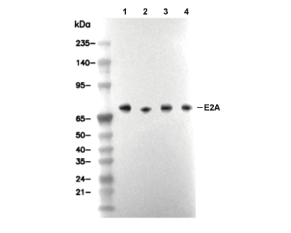 Lane 1: Jurkat, Lane 2: MOLT-4, Lane 3: K562, Lane 4: NIH/3T3
Experiment Essentials
WB
Recommended WB dilution ratio: 1:100
Recommended WB dilution ratio: 1:100
Usage Information
| Dilution |
|---|
|
| Application |
|---|
| WB, IP, IF |
| Reactivity |
|---|
| Human, Mouse, Rat |
| Source |
|---|
| Mouse Monoclonal Antibody |
| Storage Buffer |
|---|
| PBS, pH 7.2+50% Glycerol+0.05% BSA+0.01% NaN3 |
| Storage (from the date of receipt) |
|---|
| -20°C (avoid freeze-thaw cycles), 2 years |
| Predicted MW Observed MW |
|---|
| 67 kDa 63-92 kDa |
| *Why do the predicted and actual molecular weights differ? The following reasons may explain differences between the predicted and actual protein molecular weight. Post-translational modifications(e.g., phosphorylation, glycosylation); Splice variants and isoforms; Relative charge; Multimerization. |
| Positive Control | Jurkat cells; Raji cells; NIH/3T3 cells; Daudi cells; MOLT-4 cells; SUP-T1 cells; K-562 cells; HeLa cells |
|---|---|
| Negative Control |
Experimental Methods
| WB |
|---|
Experimental Protocol:
Sample preparation
1. Tissue: Lyse the tissue sample by adding an appropriate volume of ice-cold RIPA/Nuclear Lysis Buffer (containing Protease Inhibitor Cocktail),and homogenize the tissue at a low temperature. 2. Adherent cell: Aspirate the culture medium and wash the cells with ice-cold PBS twice. Lyse the cells by adding an appropriate volume of RIPA/Nuclear Lysis Buffer (containing Protease Inhibitor Cocktail) and put the sample on ice for 5 min. 3. Suspension cell: Transfer the culture medium to a pre-cooled centrifuge tube. Centrifuge and aspirate the supernatant. Wash the cells with ice-cold PBS twice. Lyse the cells by adding an appropriate volume of RIPA/Nuclear Lysis Buffer (containing Protease Inhibitor Cocktail) and put the sample on ice for 5 min. 4. Place the lysate into a pre-cooled microcentrifuge tube. Centrifuge at 4°C for 15 min. Collect the supernatant;
5. Remove a small volume of lysate to determine the protein concentration;
6. Combine the lysate with protein loading buffer. Boil 20 µL sample under 95-100°C for 5 min. Centrifuge for 5 min after cool down on ice.
Electrophoretic separation
1. According to the concentration of extracted protein, load appropriate amount of protein sample and marker onto SDS-PAGE gels for electrophoresis. Recommended separating gel (lower gel) concentration: 10%. Reference Table for Selecting SDS-PAGE Separation Gel Concentrations 2. Power up 80V for 30 minutes. Then the power supply is adjusted (110 V~150 V), the Marker is observed, and the electrophoresis can be stopped when the indicator band of the predyed protein Marker where the protein is located is properly separated. (Note that the current should not be too large when electrophoresis, too large current (more than 150 mA) will cause the temperature to rise, affecting the result of running glue. If high currents cannot be avoided, an ice bath can be used to cool the bath.)
Transfer membrane
1. Take out the converter, soak the clip and consumables in the pre-cooled converter;
2. Activate PVDF membrane with methanol for 1 min and rinse with transfer buffer;
3. Install it in the order of "black edge of clip - sponge - filter paper - filter paper - glue -PVDF membrane - filter paper - filter paper - sponge - white edge of clip"; 4. The protein was electrotransferred to PVDF membrane. ( 0.45 µm PVDF membrane is recommended ) Reference Table for Selecting PVDF Membrane Pore Size Specifications Recommended conditions for wet transfer: 200 mA, 120 min. ( Note that the transfer conditions can be adjusted according to the protein size. For high-molecular-weight proteins, a higher current and longer transfer time are recommended. However, ensure that the transfer tank remains at a low temperature to prevent gel melting.)
Block
1. After electrotransfer, wash the film with TBST at room temperature for 5 minutes;
2. Incubate the film in the blocking solution for 1 hour at room temperature;
3. Wash the film with TBST for 3 times, 5 minutes each time.
Antibody incubation
1. Use 5% skim milk powder to prepare the primary antibody working liquid (recommended dilution ratio for primary antibody 1:100), gently shake and incubate with the film at 4°C overnight; 2. Wash the film with TBST 3 times, 5 minutes each time;
3. Add the secondary antibody to the blocking solution and incubate with the film gently at room temperature for 1 hour;
4. After incubation, wash the film with TBST 3 times for 5 minutes each time.
Antibody staining
1. Add the prepared ECL luminescent substrate (or select other color developing substrate according to the second antibody) and mix evenly;
2. Incubate with the film for 1 minute, remove excess substrate (keep the film moist), wrap with plastic film, and expose in the imaging system. |
| IF |
|---|
Experimental Protocol:
Sample Preparation
1. Adherent Cells: Place a clean, sterile coverslip in a culture dish. Once the cells grow to near confluence as a monolayer, remove the coverslip for further use.
2. Suspension Cells: Seed the cells onto a clean, sterile slide coated with poly-L-lysine.
3. Frozen Sections: Allow the slide to thaw at room temperature. Wash it with pure water or PBS for 2 times, 3 minutes each time.
4. Paraffin Sections: Deparaffinization and rehydration. Wash the slide with pure water or PBS for 3 times, 3 minutes each time. Then perform antigen retrieval.
Fixation
1. Fix the cell coverslips/spots or tissue sections at room temperature using a fixative such as 4% paraformaldehyde (4% PFA) for 10-15 minutes.
2. Wash the sample with PBS for 3 times, 3 minutes each time.
Permeabilization
1.Add a detergent such as 0.1–0.3% Triton X-100 to the sample and incubate at room temperature for 10–20 minutes.
(Note: This step is only required for intracellular antigens. For antigens expressed on the cell membrane, this step is unnecessary.)
Wash the sample with PBS for 3 times, 3 minutes each time.
Blocking
Add blocking solution and incubate at room temperature for at least 1 hour. (Common blocking solutions include: serum from the same source as the secondary antibody, BSA, or goat serum.)
Note: Ensure the sample remains moist during and after the blocking step to prevent drying, which can lead to high background.
Immunofluorescence Staining (Day 1)
1. Remove the blocking solution and add the diluted primary antibody.
2. Incubate the sample in a humidified chamber at 4°C overnight.
Immunofluorescence Staining (Day 2)
1. Remove the primary antibody and wash with PBST for 3 times, 5 minutes each time.
2. Add the diluted fluorescent secondary antibody and incubate in the dark at 4°C for 1–2 hours.
3. Remove the secondary antibody and wash with PBST for 3 times, 5 minutes each time.
4. Add diluted DAPI and incubate at room temperature in the dark for 5–10 minutes.
5. Wash with PBST for 3 times, 5 minutes each time.
Mounting
1. Mount the sample with an anti-fade mounting medium.
2. Allow the slide to dry at room temperature overnight in the dark.
3. Store the slide in a slide storage box at 4°C, protected from light.
|
Biological Description
| Specificity |
|---|
| E2A Antibody [E15P20] detects endogenous levels of total E2A protein. |
| Subcellular Location |
|---|
| Nucleus |
| Uniprot ID |
|---|
| P15923 |
| Clone |
|---|
| E15P20 |
| Synonym(s) |
|---|
| Transcription factor E2-alpha; Class B basic helix-loop-helix protein 21 (bHLHb21); Immunoglobulin enhancer-binding factor E12/E47; Immunoglobulin transcription factor 1; Kappa-E2-binding factor; Transcription factor 3 (TCF-3); Transcription factor ITF-1; TCF3; BHLHB21; E2A; ITF1 |
| Background |
|---|
| E2A, also known as TCF3, is a member of the E protein family of basic helix-loop-helix (bHLH) transcription factors, alongside E2-2 and HEB. Alternative splicing of the E2A locus gives rise to the E12 and E47 isoforms, both of which contain N-terminal transactivation domains, AD1 (residues 1–100, featuring the PCET motif), AD2 (containing the LDEAI sequence), AD3 (221–300), and DES, along with a C-terminal bHLH domain (residues 547–608) responsible for dimerization and DNA binding. The bHLH domain includes a basic region (Arg547–Val559) that recognizes E-box CANNTG motifs, helix 1 (Arg560–Leu577), a loop (Lys578–Thr584), and helix 2, with Glu600 playing a key role in hydrogen bonding for homodimer formation (preferentially binding CACCTG) or heterodimerization with tissue-specific bHLH partners like MyoD (preferring CATCTG motifs). E47 homodimers are favored due to stronger interhelical contacts, whereas E12 is more prone to heterodimerization. Transcriptional activation by E2A involves AD1 inserting hydrophobic residues into KIX/TAZ2 domains of CBP/p300, which recruits histone acetyltransferases to open chromatin at immunoglobulin enhancers during B-cell lineage specification. AD2 cooperatively binds CBP KIX via the LDEAI motif, further amplifying gene activation, while the N2B motif (Pro187, Pro191, Ser192) engages the ETO NHR2 domain for corepression by recruiting HDACs, silencing genes like Nfil3 and FGFR2 in pre-B cells. AD3 interacts with TAFH4 of TAF3 in TFIID, enhancing TATA box binding and RNA Pol II recruitment for lineage-specific genes. E2A is crucial for B lymphopoiesis, activating targets such as Mb-1, Pax5, and Rag to enforce B-cell commitment and repress alternative myeloid or erythroid fates; E2A knockout mice lack B cells beyond the pro-B stage. In muscle development, E2A–MyoD/Myogenin heterodimers bind E-boxes in myogenin and desmin promoters, promoting terminal differentiation. E2A balances activation and repression, as shown by ChIP-seq: it binds directly to pro-B cell enhancers (activating) and intergenic regions (repressive). In germinal centers, E2A, together with E2-2, supports dark zone identity and somatic hypermutation via AID. E2A deficiency increases autoimmunity risk by disrupting central tolerance, whereas overexpression enhances glucocorticoid sensitivity in therapy. |
| References |
|---|
|
Tech Support
Tel: +1-832-582-8158 Ext:3
If you have any other enquiries, please leave a message.
Products are for research use only. Not for human use. We do not sell to patients.
©Copyright 2013 Selleck Chemicals. All Rights Reserved.